Cómo es el tratamiento con vitamina D contra el COVID-19 que estudia la UNCUYO

Os segredos da produção do hormônio-vitamina D – Artigo de John Cannell, MD – “Fatos não deixam de existir, apenas porque eles são ignorados.”


__


“O primeiro fato que você já conhece.   A forma ativa de vitamina D é um hormônio esteróide, e o mais potente no corpo. Os hormônios esteroides funcionam por “desmascarar” o genoma. Isto é, eles habilitam a produção de proteínas e enzimas pelo seu equipamento genético, a essência da vida. Então a forma ativa de vitamina D age habilitando a expressão genética de proteínas e enzimas cruciais para a saúde em centenas de tecidos por todo o corpo. Este fato explica por que a deficiência de vitamina D é envolvida em tantas doenças diferentes.” (…)

__

“O segundo fato mudou minha vida. Fez-me perguntar por quê?  O fato é o seguinte:  a maioria de nós produz mais ou menos 20.000 unidades de vitamina D após mais ou menos 20 minutos de sol de verão. (Para a maioria dos tipos de pele, um mínimo eritema por todo o corpo [vermelhidão leve] produzido pela luz de raios UVB resulta na produção de cerca de 20.000 unidades de colecalciferol.) Isto é mais ou menos 100 vezes mais vitamina D do que o governo diz que você precisa diariamente.” (…)

__

O terceiro fato é mais complexo e tem a ver com a regulação singular do sistema hormonal esteróide da vitamina D. Os hormônios esteroides são moléculas fabricadas a partir do colesterol que atuam agindo sobre um receptor no genoma. Os sistemas hormonais esteroides são firmemente regulados pelo organismo. Quando os níveis estão muito baixos, o corpo fabrica mais hormônios. Quando aqueles níveis estão muito altos, o corpo produz menos. Mas não com a vitamina D.


Primeiramente, diferente de outros sistemas esteroides  o sistema da vitamina D necessita de ambos, colesterol e luz solar para iniciar. O corpo não tem nenhuma maneira de obter vitamina D a menos que você entre em contato com o sol ou tome suplementos. Lembre, diferentemente de todos os outros hormônios esteroides  o corpo não pode fabricar sua própria vitamina D a partir do colesterol. Ele necessita de raios de sol também.


Claro, até mais ou menos 300 anos atrás, os humanos sempre tiveram muitos raios solares. (…)


Este complexo terceiro conjunto de fatores fortemente implica numa severa deficiência difundida entre os seres humanos modernos.  Quando os sistemas de hormônio de esteróide são ligados a pleno, sem desligamento periódico, isto normalmente significa que o corpo está sempre pedindo por mais!  Uma vez que poucos de nós vivemos desnudos sob o sol, nossos sistemas de vitamina D estão secos, nossos tanques de calcidiol estão sempre com o ponteiro na reserva, nossos tecidos estão famintos por mais desse hormônio esteróide, O MAIS POTENTE DO CORPO e, talvez por isso, as doenças de nossa civilização estejam cada vez mais disseminadas.


É por isso toda essa estapafúrdia sobre o assunto…


__

(…) Isso implica que os níveis de Vitamina D nos tecidos podem estar cronicamente esvaziados nos humanos modernos.  Além disso, nós não temos nenhum método fácil de saber se nós estamos depletados, uma vez que isso se tornou um estado humano padrão.

__

Então o que limita a quantia de calcitriol nos tecidos? A pele.

vitamin-connection

__

Trad.: José Carlos Brasil Peixoto

Você viu os recentes artigos sobre a vitamina D na Newsweek e no Boston Globe?  A manchete do artigo da Newsweek era: “Estão os americanos morrendo por falta da vitamina D?” E por que toda essa agitação sobre a vitamina D?


Todos nós sabemos que a vitamina D (colecalciferol) é crucial para sua saúde. Mas a vitamina D é realmente uma vitamina? Está presente nas comidas que os humanos normalmente consomem? Embora exista em algum percentual na gordura do peixe, a vitamina D não está em nossas dietas a não ser que os humanos artificialmente incrementem um produto alimentar, como o leite enriquecido com vitamina D. A natureza planejou que você a produzisse em sua pele, e não a colocasse direto em sua boca.


Então, seria a vitamina D realmente uma vitamina?


Diferentemente de qualquer outra vitamina, a vitamina D é realmente um pró-hormônio. O seu corpo é a única fonte do potente hormônio esteróide chamado calcitriol. Como isso naturalmente acontece? Primeiro, sua pele produz vitamina D quando a luz solar atinge uma molécula de pré-colesterol. Então seu fígado converte a vitamina D na forma de armazenamento chamado calcidiol (25-hidroxi vitamina D). O corpo armazena o calcidiol no sangue e na gordura para uso posterior.


(Seu médico pode medir o calcidiol com um exame de sangue para descobrir se você apresenta deficiência de vitamina D. Os níveis ideais de calcidiol [25-hidroxi vitamina D] estão entre 35-65 ng/ml [87-162 nm/L], ao longo do ano.)

Se você tiver suficiente calcidiol em seu sangue, então as “coisas realmente acontecem”. Um pouco de calcidiol vai para os rins para ajudar a manter os níveis de cálcio no sangue, mas o mais importante acontece nos seus tecidos. Os tecidos por toda parte do seu corpo convertem calcidiol em calcitriol. O calcitriol, ou vitamina D ativa, é o hormônio esteróide mais potente do corpo humano. É ativo em quantidades medidas em pico gramas ou 1/1.000.000.000.000 de um grama.


Como os todos os hormônios esteroides, o calcitriol funciona ligando seus genes. Isto é, em centenas de tecidos por todo o seu corpo, o calcitriol expõe seu genoma! Ele dá o sinal para seus genes produzirem centenas de enzimas e proteínas cruciais para manter a saúde e lutar contra doenças.


Obtendo Sua Porção de Vitamina D


Tudo isso acontece, só se você conseguir suficiente vitamina D a partir dos raios do sol ou de suplementos. Se você é um dos que evitam totalmente o sol, pesquisas recentes indicam que você precisa mais ou menos 4.000 unidades de vitamina D em um dia! Então você não vai conseguir suficiente vitamina D a partir do leite (a menos que você beba 40 copos por dia) ou de um multi-vitamínico (a menos que você ingira mais ou menos 10 tabletes por dia), aliás, nada disso é recomendado.


Se você não conseguir a vitamina D do modo como a Mãe Natureza pretendeu, a partir do raio do sol, você precisa tomar suplementos de vitamina D. Muitos de nós conseguimos muito mais vitamina D dos raios solares do que nós imaginamos, mas a maioria tem uma necessidade de mais ou menos 2.000 unidades extras por dia. Cada vez mais a Internet e lojas de suplementos alimentares e de saúde vendem vitamina D.


Tenha certeza de que a vitamina D que você compra é puro colecalciferol. Não compre uma preparação adicionada com vitamina A. Apenas puro colecalciferol.


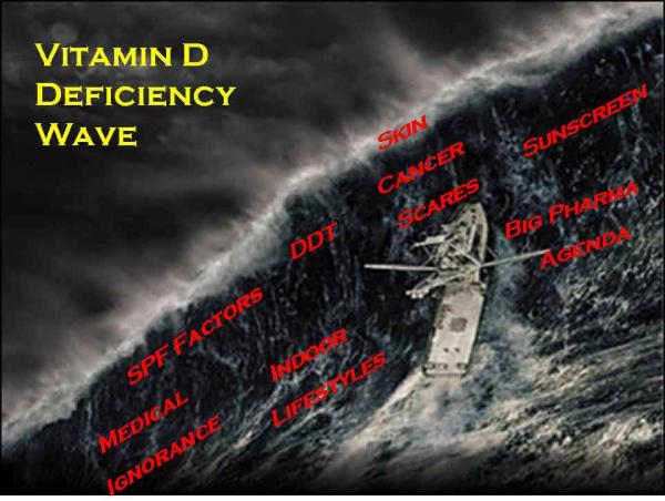
Muitas das doenças de civilização moderna – câncer, doença de coração, diabete, hipertensão, doença periodental, depressão e até obesidade – estão agora claramente associados com a deficiência de vitamina D. Mas uma associação não é o mesmo que uma relação de causa e efeito. A deficiência de vitamina D causa muitos casos dessas doenças da civilização moderna? Nós apenas não sabemos. Nós precisamos dos Institutos Nacionais de Saúde para financiar mais pesquisa em vitamina D. Até agora, porém, eles recusaram.


Se você quiser entender a vitamina D, você precisa reconhecer três fatos que têm sido geralmente ignorado por quase todos exceto alguns cientistas da vitamina D.  Aldous Huxley uma vez disse, “Fatos não deixam de existir, apenas porque eles são ignorados.”  Dois destes fatos ignorados são questões simples e um terceiro é mais complexo.


O Hormônio Esteróide


O primeiro fato que você já conhece.  A forma ativa de vitamina D é um hormônio esteróide, e o mais potente no corpo. Os hormônios esteróides funcionam por “desmascarar” o genoma. Isto é, eles habilitam a produção de proteínas e enzimas pelo seu equipamento genético, a essência da vida. Então a forma ativa de vitamina D age habilitando a expressão genética de proteínas e enzimas cruciais para a saúde em centenas de tecidos por todo o corpo. Este fato explica por que a deficiência de vitamina D é envolvida em tantas doenças diferentes.


O segundo fato mudou minha vida. Fez-me perguntar por quê? O fato é o seguinte: A maioria de nós produz mais ou menos 20.000 unidades de vitamina D após mais ou menos 20 minutos de sol de verão. (Para a maioria dos tipos de pele, um mínimo eritema por todo o corpo [vermelhidão leve] produzido pela luz de raios UVB resulta na produção de cerca de 20.000 unidades de colecalciferol.) Isto é mais ou menos 100 vezes mais vitamina D do que o governo diz que você precisa diariamente.


Pergunte a si mesmo: por quê? Por que os seres humanos fariam tanta vitamina D, com tanta rapidez? Eu pensei sobre isto, estudei livros de ensino, pesquisei na literatura médica, perguntei a todos os peritos, e dediquei o resto de minha vida profissional a fazer outras pessoas a se perguntarem “por quê?” Por que nós teríamos um sistema hormonal esteróide que faz tanto substrato com tanta rapidez?


A única resposta que qualquer um pode apresentar é: “Provavelmente seja por uma boa razão.” A ciência não sabe por quê. Os biólogos sabem que a natureza não projeta sistemas tão complexos quanto o sistema hormonal esteróide da vitamina D sem alguma razão. A ciência médica simplesmente não sabe por que nós temos a capacidade para produzir tanta vitamina D tão depressa.


Se você pensar sobre isso por um tempo razoável, você também concluirá que é provavelmente para uma boa razão. Embora nós não saibamos por que, alguns cientistas têm tentado descobrir porque, e perdem o fôlego nas explosivas possíveis implicações.


Este segundo fato também diz a você algo sobre a condição humana normal – e o atual desvio de conduta. Antes de nós começarmos a viver em edifícios e carros, vestindo roupas protetoras contra o sol e besuntando em bloqueadores solares, nós lavrávamos e caçamos. E antes disso, nós procurávamos por alimentos, desnudos sob o sol subequatorial africano por mais de um milhão de anos.


Quanta vitamina D nós obtínhamos então? Muita.


Nós começamos a movermos para os interiores durante a revolução industrial e agora o movimento está quase completo.  Alguns de nós ficamos por dias, semanas, ou até meses sem deixar que um único raio de sol atinja nossa pele e produza vitamina D. Se nós formos ao sol, nossos dermatologistas nos repreendem.   Tanto faz se é bom ou ruim, esta forma de existência é aberrante para a espécie. A “moderna” evitação ao sol é um erro de conduta para o homo sapiens.


Uma vez que nós produzimos mais ou menos 20.000 unidades de vitamina D com alguns minutos de raio de sol (talvez 10.000 unidades após nossa pele ficar bronzeada), foi assim que o ser humano fez para ter muita vitamina D a cada dia, até muito recentemente. Agora, a maior parte de nós obtém muito pouco. Isto é simplesmente um desvio.


Controlando Natureza?


O terceiro fato é mais complexo e tem a ver com a regulação singular do sistema hormonal esteróide da vitamina D. Os hormônios esteroides são moléculas fabricadas a partir do colesterol que atuam agindo sobre um receptor no genoma. Os sistemas hormonais esteroides são firmemente regulados pelo organismo. Quando os níveis estão muito baixos, o corpo fabrica mais hormônios. Quando aqueles níveis estão muito altos, o corpo produz menos. Mas não com a vitamina D.


Primeiramente, diferente de outros sistemas esteroides  o sistema da vitamina D necessita de ambos, colesterol e luz solar para iniciar. O corpo não tem nenhuma maneira de obter vitamina D a menos que você entre em contato com o sol ou tome suplementos. Lembre, diferentemente de todos os outros hormônios esteroides  o corpo não pode fabricar sua própria vitamina D a partir do colesterol. Ele  necessita de raios de sol também.


Claro, até mais ou menos 300 anos atrás, os humanos sempre tiveram muitos raios solares.


Lembre, a ação real está nos tecidos.  O sistema de vitamina D autócrino (para a própria célula) e parácrino (para as células vizinhas) parecem estarem ligados a pleno o tempo todo. (Em termos científicos, a constante de Michaelis Menton nunca é alcançada plenamente até que ocorra o pleno equilíbrio das taxas de concentração dos substratos fisiológicos de ambas, a produção de calcidiol do fígado e da produção de calcitriol dos tecidos.)


O sistema direto de retroalimentação negativa (direct negative feedback) não parece estar operando em níveis fisiológicos para ambas as produções de calcidiol no fígado e calcitriol nos tecidos. Isso implica que os níveis nos tecidos podem estar cronicamente esvaziados nos humanos modernos. Além disso, nós não temos nenhum método fácil de saber se nós estamos depletados, uma vez que isso se tornou um estado humano padrão.


Se a produção de tecido de calcitriol está ligada a pleno, o tempo todo, o que previne a toxicidade da vitamina D nos humanos que vivem sob o sol?  Primeiro muito da vitamina D você produz é excretado pela bílis. O mesmo pode ser verdade para muito do calcidiol que seu fígado produz. Além disso, existem numerosos outros metabólitos da vitamina D. Então, apenas mais ou menos 1/1000 de seu calcidiol é transformado em calcitriol.  Dito isso, a produção nos tecidos de calcitriol está ainda correndo a pleno sob concentrações normais do substrato calcidiol.


Então o que limita a quantia de calcitriol nos tecidos? A pele.


Depois de você produzir mais ou menos 20.000 unidades, os raios de sol começam a destruir vitamina D na pele. Em outras palavras, a mesma luz solar que produz vitamina D é a primeira a iniciar o seu processo de degradação. A produção equivale à destruição.


Como a produção de calcitriol nos tecidos e a criação de calcidiol no fígado sempre funcionam abaixo de sua capacidade bioquímica, isso significa que o processo limitador das taxas do hormônio esteroide mais potente do corpo humano parece recair sobre a pele.  De certo modo, isso recai sobre seu comportamento, sua escolha em andar ao sol – ou não. Isto é biologicamente inigualável para qualquer um de todos os hormônios esteroides.]


Este complexo terceiro conjunto de fatores fortemente implica numa severa deficiência difundida entre os seres humanos modernos.  Quando os sistemas de hormônio de esteróide são ligados a pleno, sem desligamento periódico, isto normalmente significa que o corpo está sempre pedindo por mais!  Uma vez que poucos de nós vivemos desnudos sob o sol, nossos sistemas de vitamina D estão secos, nossos tanques de calcidiol estão sempre com o ponteiro na reserva, nossos tecidos estão famintos por mais desse hormônio esteróide, O MAIS POTENTO DO CORPO, e, talvez por isso, as doenças de nossa civilização estejam cada vez mais disseminadas.


É por isso toda essa estapafúrdia sobre o assunto…


Saiba mais sobre o assunto no site:

http://www.vitamindcouncil.org

Conheça melhor o autor:

http://www.vitamindcouncil.org/cannellBiography.shtml

Deficiência de Vitamina D: Um importante fator de risco para a doença cardiovascular

__

A vitamina D não é uma “vitamina”. Seu produto metabólico, calcitriol , é realmente um hormônio seco esteróide que é a chave que abre pontos de ligação do genoma humano. O genoma humano contém mais de 2.700 sítios de ligação para o calcitriol, que estão perto dos genes envolvidos em PRATICAMENTE TODAS AS DOENÇAS importantes conhecidas dos seres humanos.

genoma

__

Conforme estudos recentemente publicados, a deficiência de Vitamina D   [que  não é vitamina  e  sim  um hormônio  esteroide ASSOCIADO à SAÚDE  de todas as pessoas],  que é tradicionalmente associada com fraqueza óssea ou muscular,  também aumenta o risco da doença cardiovascular. Um crescente número de evidências relaciona baixos níveis de Vitamina D a fatores comuns da doença cardiovascular, como por exemplo, hipertensão arterial, diabetes, aterosclerose, além de outros eventos cardiovasculares maiores que incluem acidente vascular cerebral, insuficiência cardíaca e morte súbita por origem cardíaca. Mundialmente a prevalência da deficiência de vitamina D entre idosos é de quase 50%.

Resultados dos estudos sobre vitamina D e doença cardiovascular

Tem sido demonstrado que a deficiência de Vitamina D leva a diminuição na contratilidade cardíaca, tônus vascular, volume do colágeno cardíaco e desenvolvimento do tecido cardíaco, existindo uma crescente evidência vinculando resultados do tratamento com vitamina D para a melhoria da sobrevida na hemodiálise em pacientes no estágio final da doença renal, e melhoria na função cardíaca (1, 2).

Baixos níveis de vitamina D no plasma têm sido relacionados como um fator contributivo para a patogênese da insuficiência cardíaca congestiva (3), prevalente disfunção miocárdica, óbitos por insuficiência cardíaca congestiva, e morte cardíaca súbita (4).

Indivíduos com baixos níveis de vitamina D têm maior incidência de pressão alta, diabetes, e elevação nos triglicérides do que aqueles com mais altos níveis de vitamina D no plasma (5).

Estudos epidemiológicos recentes também têm vinculado a deficiência de vitamina D com um aumentado risco de eventos adversos cardiovasculares maiores (6). Um estudo envolvendo profissionais da saúde mostrou um risco 2 vezes maior de enfartes do miocárdio em indivíduos com deficiência D comparados com aqueles dentro de uma faixa normal (7). Similarmente um estudo prospectivo recente de coortes mediu os níveis de vitamina D em 3.258 pacientes adultos, os quais estavam sendo submetidos a cateterização cardíaca eletiva. Durante o seguimento médio de 7.7 anos, os indivíduos no mais baixo quartíl de 25-hydroxyvitamin D no soro tiveram um risco ajustado de 2 vezes no aumento de risco de óbito, especialmente óbito por doença cardiovascular, comparados com aqueles no mais alto quartil de vitamina D (8).

Baixos níveis de vitamina D aumentam o risco de eventos cardiovasculares entre pacientes com hipertensão, mas não entre aqueles sem hipertensão (6). Mulheres jovens com baixos níveis de vitamina D no plasma têm um aumentado risco para o desenvolvimento de hipertensão (9).

Níveis maiores de calcificação coronária são vistos naqueles pacientes com maior deficiência de vitamina D (10, 11).

A deficiência de vitamina D pode se estender a doenças vasculares além das artérias coronárias. Recente estudo determinou que quanto maior a deficiência de vitamina D, mais placa aterosclerótica era encontrada na carótida, através de avaliação por ultrasonografia. A espessura da íntima-medial da carótida é considerada uma medida substituta válida para a avaliação da aterosclerose coronária. Esse estudo concluiu que os baixos níveis de vitamina D encontrados no sangue representam um forte e independente preditor da aterosclerose. Também mostrou que a deficiência severa de vitamina D foi o dobro em diabéticos do que em não diabéticos (12).

Pessoas com pouca ou nenhuma função renal freqüentemente têm muitos baixos níveis de vitamina D, já que o rim é requerido para essa ativação (13). Estudo envolvendo pacientes com doença renal grave em diálise peritoneal demonstrou que o tratamento para corrigir a deficiência de vitamina D conseguiu uma dramática redução em enfartes e óbitos por doença cardíaca (14). Outro estudo revisando as evidências, tanto da ciência básica quanto de estudos clínicos, apoiou um possível papel protetor da vitamina D, além de seus efeitos no metabolismo mineral, sugerindo a necessidade para uma avaliação contínua do papel da vtamina D na saúde cardiovascular da população de pacientes com doença renal crônica (15).

Enfim, a restauração da vitamina D aos níveis normais pode ajudar a reduzir inflamação, a normalização da pressão sangüínea, e melhora da sensibilidade à insulina – fatores que podem reduzir o risco cardíaco (16, 17, 18, 19).

Estudo recentemente publicado no Jornal do Colégio Americano de Cardiologia emitiu recomendações práticas para a triagem e tratamento de pacientes com baixos níveis de vitamina D, especialmente naqueles com fatores de risco para doenças cardíacas e diabetes, ressaltando que a vitamina D é fácil de conseguir e sua suplementação é simples, segura e de baixo custo (20).

Fontes de vitamina D

A pele humana é rica em pré-vitamina D latente, a qual é ativada pela luz do sol. A vitamina D não é contida em um grau significante nos alimentos e nem deveria ser obtida através de consumo oral. Algumas das fontes naturais de vitamina D incluem salmão, sardinhas, óleo de fígado de bacalhau, e de alimentos fortificados com vitamina D, incluindo leite e alguns cereais. A vitamina D pode também ser fornecida na forma de suplementos.

As pessoas deveriam obter vitamina D através da exposição ao sol. Entretanto, através dos séculos as pessoas têm migrado para lugares mais frios e privados de climas mais ensolarados, trabalhando internamente em escritórios ou fábricas, fazendo viagens de automóvel, exercícios em ginásios ao invés de ao ar livre, e vestindo roupas que cobrem praticamente 95% da superfície do corpo. De fato, mesmo adultos vivendo em lugares ensolarados são passiveis de deficiência de vitamina D, pelo menos durante uma parte do ano. Assim o consumo oral de vitamina D se tornou necessário tendo em vista que os seres humanos desenvolveram estilos de vida envolvendo menos e menos exposição ao sol. Adicionando-se a isso a fobia promovida pelos dermatologistas que aconselham a população de que exposição ao sol causa câncer de pele, com muitas pessoas privando-se do sol e, portanto incapazes de ativar a vitamina D (21).

A teoria de Carl J Reich sobre a deficiência de cálcio e vitamina D

Há cerca de 53 anos Carl Reich acreditava que muitos dos sintomas e doenças da civilização poderiam ser atribuídos à deficiência crônica de cálcio e vitamina D, criada por defeitos específicos no estilo de vida, incluindo dieta, podendo levar à acidose.

No início de sua prática clínica nos anos 50 ele acompanhou diversos pacientes exibindo todos os sinais físicos e sintomas de um sistema nervoso autônomo sobre-estimulado os quais muito freqüentemente eram classificados como de origem psicossomática, como por exemplo: fadiga crônica, fraqueza física, ansiedade, distúrbios do sono, dores de cabeça, câimbras, etc…

Reich descobriu que seus pacientes saudáveis tinham leituras na saliva, feitas através de papel litmus, com medidas de neutro para alcalino de 7,0 para 7,5 pH que ele acreditava fielmente representassem o pH do sangue e tecidos. Enquanto isso os pacientes não saudáveis tinham leituras de saliva ácida mostrando evidência de um pH ácido no sangue na direção de ou abaixo de 6,5.

Dentro de poucas semanas ou meses de dieta com vitaminas e minerais e mudanças suplementares o pH ácido na saliva dos pacientes tratados podiam retornar a leituras mais normais de neutro para alcalino, de 7 para 7,5 pH, acompanhadas pelo desaparecimento de sintomas (22)

Referências

  1. Zittermann A. Vitamin D and disease prevention with special reference to cardiovascular disease. Prog Biophys Mol Biol. 2006 Sep;92(1):39-48.

  2. Achinger SG, Ayus JC. The role of vitamin D in left ventricular hypertrophy and cardiac function. Kidney Int Suppl. 2005 Jun;(95):S37-S42.

  3. Zittermann A, Schleithoff SS, Tenderich G, Berthold HK, Korfer R, Stehle P. Low vitamin D status: a contributing factor in the pathogenesis of congestive heart failure? J Am Coll Cardiol. 2003 Jan 1;41(1):105–12.

  4. Pilz S, Marz W, et al. Association of vitamin D deficiency with heart failure and sudden cardiac death in a large cross-sectional study of patients referred for coronary angiography. J Clin Endocrinol Metab, Oct 2008, 93(10): 3927-3935

  5. Martins D, Wolf M, Pan D, et al. Prevalence of cardiovascular risk factors and the serum levels of 25-hydroxyvitamin d in the United States: data from the third national health and nutrition examination survey. Arch Intern Med. 2007 Jun 11;167(11):1159-65.

  6. Wang T, Pencina MJ, Booth SL, et al. Vitamin D deficiency and risk of cardiovascular disease. Circulation, 2008 Jan 29;117(4):503-11.

  7. Giovannuci E, Liu Y, Hollis BW, Rimm EB. 25-Hydroxyvitamin D and risk of myocardial infarction in men: a prospective study. Arch Intern Med 2008;168:1174-80

  8. Dobnig H, Pilz S, Schanagl H et al. Independent association of low serum 25-Hydroxyvitamin D  and 1,25-Hydroxyvitamin D levels with all cause and cardiovascular mortality. Arch Intern Med 2008;168:1340-9

  9. Forman JP, Curhan GC, Taylor EM. Plasma 25-Hydroxyvitamin D levels and risk of hypertension among young women. Hypertension. 2008, 52:828

  10. Watson KE, Abrolat ML, Malone LL, et al. Active serum vitamin D levels are inversely correlated with coronary calcification. Circulation. 1997 Sep 16;96(6):1755-60.

  11. Zittermann A, Schleithoff SS, Koerfer R. Vitamin D and vascular calcification. Curr Opin Lipidol. 2007 Feb;18(1):41-6.

  12. Targher G, Bertolini L, Padovani R, et al. Serum 25-hydroxyvitamin D3 concentrations and carotid artery intima-media thickness among type 2 diabetic patients. Clin Endocrinol (Oxf). 2006 Nov;65(5):593-7.

  13. Levin A, Li YC. Vitamin D and its analogues: do they protect against cardiovascular disease in patients with kidney disease? Kidney Int. 2005 Nov;68(5):1973-81.

  14. Taskapan H, Ersoy FF, Passadakis PS, et al. Severe vitamin D deficiency in chronic renal failure patients on peritoneal dialysis. Clin Nephrol. 2006 Oct;66(4):247-55.

  15. Shoji T, Shinohara K, Kimoto E, et al. Lower risk for cardiovascular mortality in oral 1alpha-hydroxy vitamin D3 users in a haemodialysis population. Nephrol Dial Transplant. 2004 Jan;19(1):179-84.

  16. Pfeifer M, Begerow B, Minne HW, Nachtigall D, Hansen C. Effects of a short-term vitamin D(3) and calcium supplementation on blood pressure and parathyroid hormone levels in elderly women. J Clin Endocrinol Metab. 2001 Apr;86(4):1633-7.

  17. Lind L, Hanni A, Lithell H, et al. Vitamin D is related to blood pressure and other cardiovascular risk factors in middle-aged men. Am J Hypertens. 1995 Sep;8(9):894-901.

  18. Timms PM, Mannan N, Hitman GA, et al. Circulating MMP9, vitamin D and variation in the TIMP-1 response with VDR genotype: mechanisms for inflammatory damage in chronic disorders? QJM. 2002 Dec;95(12):787-96.

  19. Zittermann A. Vitamin D and disease prevention with special reference to cardiovascular disease. Prog Biophys Mol Biol. 2006 Sep;92(1):39-48.

  20. Lee JH, O’Keefe JH, et al. Vitamin D deficiency, an important, common, and easily treatable cardiovascular risk factor?. J Am Coll Cardiol 2008; 52: 1949-1956

  21. William Davis,, Vidamin D’s crucial role in cardiovascular protection, LE Magazine, September 2007.

  22. Calcium and Vitamin D Deficiency. The clinical work and theory of Carl J. Reich, MD. Publicado em 1995 pela “Foundation for Erradication of Rheumatoid Disease”, e disponível em http://www.arthritistrust.org/Articles/Calcium%20and%20Vitamin%20D%20Deficiency.pdf

Fonte:  http://www.infarctcombat.org/polemica-47/icem.html

Obesidade pode levar à deficiência de vitamina D, mas o contrário não ocorre


__


Pesquisadores  estimam  que  cada  10%  de  aumento  no  IMC  leva  a  4,2%  de  queda  na  concentração  de vitamina D


Obesidade-e-Diabetes


A obesidade pode levar à deficiência de vitamina D, mas a deficiência de vitamina D não leva à obesidade. Essa é a principal conclusão de um amplo estudo que analisou dados de 42.024 participantes de 21 pesquisas, publicado nesta terça-feira no periódico científico Plos Medicine.

English: A Collection of Articles on Disease M...

English: A Collection of Articles on Disease Mongering in PLoS Medicine Español: Portada del monográfico publicado en Public Library of Science – Medicine sobre promoción de enfermedades (Photo credit: Wikipedia)

Para os autores, a descoberta tem grande importância para a saúde pública,  uma vez que a obesidade vem crescendo em todo o mundo, o que indica que a deficiência de vitamina D também pode aumentar.


A vitamina D é produzida pela pele após a exposição ao sol, mas também pode ser obtida através da alimentação. Ela é encontrada principalmente em leite e derivados, além de peixes oleosos, como cação e salmão, ricos em ômega 3. A falta dessa vitamina pode causar, além de problemas ósseos, alterações na liberação de insulina e no controle da pressão arterial, por exemplo.


O estudo relacionou os principais genes ligados à obesidade com aqueles referentes à vitamina D, e a associação entre o aumento do índice de massa corporal (IMC – calcule aqui seu IMC) e a redução da vitamina D se mostrou muito consistente.  Os pesquisadores  estimam que cada 10% de aumento no IMC leva a 4,2% de queda na concentração de vitamina  D  mas,  por outro lado, a deficiência de vitamina D não exerce influência sobre o IMC.


“Nosso estudo sugere que uma intervenção a nível populacional para reduzir a obesidade poderia levar a uma redução na prevalência de deficiência de vitamina D”, afirmam os autores.


Fonte: http://veja.abril.com.br/noticia/saude/obesidade-pode-levar-a-deficiencia-de-vitamina-d-mas-o-contrario-nao-ocorre


__


Cientistas convocam para uma Ação de Saúde Pública tendo como modelo o uso do Hormônio-Vitamina D

__

CIENTISTAS CONVOCAM PARA UMA AÇÃO D*  

[*PROJETO QUE TEM O PROPÓSITO DE SERVIR COMO UM MODELO DE SAÚDE PÚBLICA COM A VITAMINA D] 

A DEFICIÊNCIA DE VITAMINA D É EPIDÊMICA
__

Sobre o assunto, assista também: Vitamina D3 – 10.000 UI diárias é vital para à saúde

__
Scientists’ Call to D*action
The Vitamin D Deficiency Epidemic

vita d1

40-75% of the world’s population is vitamin D deficient.

The causal link between severe vitamin D deficiency and rickets or the bone disease of osteomalacia is overwhelming, while the link between vitamin D insuffiency and osteoporosis with associated decreased muscle strength and increased risk of falls in osteoporotic humans is well documented by evidencebased intervention studies.

There are newly appreciated associations between vitamin D insufficiency and many other diseases, including tuberculosis, psoriasis, multiple sclerosis, inflammatory bowel disease, type-1 diabetes, high blood pressure, increased heart failure, myopathy, breast and other cancers which are believed to be linked to the non-calcemic actions of the parent vitamin D and its daughter steroid hormone. Based on the evidence we now have at hand, action is urgent.

It is projected that the incidence of many of these diseases could be reduced by 20%-50% or more, if the occurrence of vitamin D deficiency and insufficiency were eradicated by increasing vitamin D intakes through increased UVB exposure, fortified foods or supplements. The appropriate intake of vitamin D required to effect a significant disease reduction depends on the individual’s age, race, lifestyle, and latitude of residence. The latest Institute of Medicine (IOM) report, 2010, indicates 10,000 IU/day is considered the NOAEL (no observed adverse effect level). 4000 IU/day can be considered a safe upper intake level for adults aged 19 and older.

It is well documented that the darker the skin, the greater the probability of a vitamin D deficiency. Even in southern climates, 55% of African Americans and 22% of Caucasians are deficient.

More than 1 billion people worldwide are affected at a tremendous cost to society.

A Scientists’ Call to Action has been issued to alert the public to the importance to have vitamin D serum levels between 40 and 60 nanograms/milliliter (100-150 nanomoles/liter) to prevent these diseases. Implementing this level is safe and inexpensive.

The benefit of an adequate vitamin D level to each individual will be better overall health and a reduction in illnesses and, ultimately, a significant reduction in health care costs. The benefit of adequate vitamin D levels to society/businesses is a more productive workforce and, lower health care costs.

The D*action project has as its purpose to serve as a model for public health action on vitamin D. It is a test bed for techniques, and for providing outcome evaluation at a community level.

http://www.grassrootshealth.net/epidemic

A Revista Veja desta semana tem como matéria de capa o assunto Vitamina D. Ed. 2.304, de 16 de janeiro de 2013: “O que você não sabe sobre a Vitamina do Sol. Ela continua a surpreender a medicina com novos efeitos benéficos.”

veja

Para receber as informações mais atualizadas sobre este hormônio-vitamina D, cadastre-se, curtindo a página:

Vitamina D é um hormônio vital para preservação e recuperação da saúde

https://www.facebook.com/VitaminaD.HormonioVital

__

Na COLUNA DA ESQUERDA deste site está situado em último lugar a categoria “VITAMINA D”.  Entrem ali e terão acesso às principais publicações, vídeos e programas feitos sobre esta vitamina-hormônio.  Ou apenas cliquem no link que dá acesso direto a todas elas:

 https://biodireitomedicina.wordpress.com/category/vitamina-d/

__

No meu canal do YouTube, todo o material de áudio, vídeos e programas sobre Vitamina D3 podem ser acessados neste endereço:

Vitamina D3 – 10.000 UI diárias é vital para preservar à saúde

https://www.youtube.com/playlist?list=PL301EAE2D5602A758

Celso Galli Coimbra – OABRS 11352
cgcoimbra@gmail.com
 

Temporada de nascimento. Baixa Exposição à Luz Solar – deficiência de Vitamina D é associada com risco elevado de esquizofrenia

__

Este trabalho refere-se a períodos de estações do hemisfério norte.  No hemisférios sul os meses correspondentes são opostos.
Schz.month2

Risco relativo de esquizofrenia dependendo do mês de nascimento

De acordo com um artigo da revista New Scientist, uma pesquisa sugere que pessoas que desenvolvem esquizofrenia na Europa e América do Norte são mais prováveis de terem nascido no inverno e começo da primavera (Fevereiro e Março no Hemisfério Norte).

Em outras palavras, os indivíduos nascidos durante esses meses tem uma ligeira maior incidência que a média de desenvolver esquizofrenia, enquanto que indivíduos nascidos em Agosto e Setembro tem uma incidência ligeiramente menor que a média. Parece haver uma diferença de 10% no risco de desenvolver esquizofrenia entre os meses de nascimento de alto risco (Inverno e Primavera) e de baixo risco.

Uma possível razão que os pesquisadores acreditam que possa explicar essa temporada de risco de esquizofrenia é a associação entre os nascimentos ocorridos no inverno/primavera e a esquizofrenia possivelmente causada pela exposição à luz solar. Uma carência de luz solar (por exemplo, durante os dias mais curtos do inverno) poderia levar à deficiência de vitamina D, que os cientistas acreditam poder alterar o desenvolvimento do cérebro da criança no útero da mãe e após o nascimento.

O que fazer: Para um risco reduzido de esquizofrenia, a mãe poderá se certificar de receber pelo menos o dobro da dose recomendada de vitamina D regularmente antes e durante a gravidez. Em um recente estudo sobre deficiência de vitamina D durante a gravidez, a Dra. Lisa Badner observou que “Enquanto várias vitaminas pré-natais contem 400 unidades internacionais (IU) de vitamina D, absorver por volta de 1.000 seria o preferível“. A vitamina D pode ser adquirida como suplemento nutricional que não precise de prescrição médica, ou pode ser incluído na compra em supermercados de leites e sucos de laranja.

Um casal planejando uma criança também pode tentar marcar o parto para um período fora do intervalo entre “inverno e primavera” (intervalos que foram implicados como mais suscetíveis a um número maior que a média de pessoas nascidas que futuramente desenvolvem esquizofrenia). Pesquisas sugerem que os meses de nascimento que tem menor risco são Julho até Outubro.

Fonte (sem tradução): http://www.schizophrenia.com/prevention/season.html (tem mais nesse link, um deles que sugere que o nível adequado de vitamina D seria de 2000 IU durante o primeiro ano de vida).

E que o protetor solar não causa deficiência de vitamina D, apenas reduz o risco real de câncer de pele, sendo que 90% desses são causados por muita exposição ao Sol com a pele desprotegida. Outro problema associado com falta de vitamina D na infância é o raquitismo – amolecimento dos ossos.

Risk factors in schizophrenia. Season of birth, gender, and familial risk.

The risk for schizophrenia among first-degree relatives of schizophrenic probands obtained from an epidemiological sample using family history methods was examined to determine whether month of birth of the proband was associated with familial risk.

The results of this study of the first-degree relatives of 106 female schizophrenics and 275 male schizophrenics suggested that the relatives of probands born in the months February to May had the highest risk, although the association between month of birth and familial risk among the male probands was present only for those relatives who had onset of schizophrenia before the age of 30.

Prenatal

Um número de fatores durante o desenvolvimento do feto pode resultar num pequeno aumento no risco de esquizofrenia futuramente na vida incluindo: hipoxia (deficiência de oxigênio nos tecidos orgânicos), e infecção, estresse ou má nutrição da mãe.

Pessoas diagnosticadas com esquizofrenia são mais suscetíveis a terem nascido no inverno ou primavera (pelo menos no hemisfério norte). Isso pode se dever aos níveis elevados de exposições virais no útero.

Que fatores ambientais são importantes?

A meta-analysis has shown that patients with schizophrenia are more likely to have experienced obstetric complications, in particular premature birth, low birth weight, and perinatal hypoxia.

Eles também são mais prováveis de terem nascido no fim do inverno e começo da primavera, possivelmente refletindo exposições virais intra-uterinas. Esses riscos ambientais prematuros aparentemente tem um efeito sutil no desenvolvimento cerebral.

In adulthood different environmental stressors act — including social isolation, migrant status, and urban life — and this remains the case even when life events attributable to the incipient psychosis itself are excluded.

The way parents raise their children does not seem to have a major impact on future vulnerability, but families do have an important part to play in the course of the illness; patients with supportive parents do much better than those with critical or hostile ones.

Collectively, these risk factors point to an interaction between biological, psychological, and social risk factors that drive increasingly deviant development and finally frank psychosis.

Fontes:

http://www.ncbi.nlm.nih.gov/pmc/articles/PMC1914490/

Seu filho recebe Vitamina D suficiente? Does your child get enough vitamin D?

__

vitaminaD2

Vitamin D is an essential nutrient that a growing child’s body needs to help absorb calcium and phosphorus, which are important for healthy bones and teeth.

Vitamin D mostly comes from summer sunlight as well as certain foods.

Not getting enough vitamin D has led to some children in the UK developing the rare bone disease rickets. Rickets was common during Victorian times but mostly disappeared in the 1940s.

Even if children get enough calcium from their diet, including through milk and yoghurt, without enough vitamin D, calcium cannot be properly absorbed.

Vitamin D also regulates cell growth, neuromuscular and immune function and reduction of inflammation.

Vitamin D from the sun

The British climate may be responsible for some vitamin D deficiency. 90% of vitamin D intake comes from exposure of the skin to sunlight. Between October and March, there’s not enough UVB radiation in autumn, winter and early spring sunlight.

The body can store up vitamin D over the summer months, but the amount of UVB radiation necessary to produce vitamin D also depends on a child’s skin colour.

Those with lighter complexions need less sun exposure than those with darker skin. Some children may have less exposure to the sun because their skin is covered for cultural reasons. Others may miss out because of having to stay indoors or in hospital due to medical conditions.

A balance is important between getting children out on the sun enough for their vitamin needs while making sure their delicate skin is protected from sun burn or over exposure.

Experts advise exposing the skin to regular, short periods of sun during the summer months, without sunscreen, which blocks UVB rays.  There is no official recommendation for time limits, but a short period unprotected in the sun can be around 10 to 15 minutes for most children. After that, the normal sun protection measures are needed. These include covering up and using sunscreen with a sun protection factor (SPF) of at least 15.

Vitamin D from food

Vitamin D occurs naturally in some foods, including oily fish and eggs, and is added to some foods such as spreads and breakfast cereal.

Breastfed babies get their vitamin D from their mother’s breast milk, so it is also important for breastfeeding women to get enough vitamin D themselves. Pregnantand breastfeeding women are advised to take a daily supplement containing 10 micrograms of vitamin D for the needs of the mother and growing baby.

Vitamin D from supplements

The Department of Health recommends supplement drops containing vitamin D for babies and children from six months to five years old.

This will give them their daily requirement of 7-8.5 micrograms of vitamin D.

Infant formula is fortified with vitamin D, so babies who have formula won’t need vitamin drops until they have less than 500ml (around a pint) of infant formula a day.
Check with your health visitor or GP if you have concerns.

If a breastfeeding mother didn’t have vitamin D supplements throughout her pregnancy, a health visitor may say the baby needs vitamin drops from one month old.

Women and children may qualify for free supplements containing vitamin D under the Healthy Start programme. Age-appropriate vitamin drops are also available from pharmacies and supermarkets

Fonte: http://www.webmd.boots.com/children/child-vitamin-d


Are you one of those who believe plain milk is enough for your child’s nutritional needs? Have you ever thought about your child’sVitamin D levels? These findings are an eye-opener

Mark ‘yes’ if these situations are relevant to your child:

1. Your child barely plays out in the sun.

2. Off late your child tends to get tired easily & seems listless at times.

3. Your child complains of pain in his joints without reason.

If your answers are predominantly ‘yes’, then this recent 11-city survey conducted by Ipsos Research will be an eye-opener for you. The purpose was to understand the level of awareness about Vitamin D among mothers – whether they considered it important enough for their child, the misconceptions about it and the harm that would befall the child if they are found to be Vitamin D deficient. The response from the 2000 mothers who were spoken to for the survey, threw up some compelling insights.

First the good news. General awareness levels among the mothers were high: most of them correctly mentioned sunlight as a source of Vitamin D. They were aware that lack of this vitamin would result in weak bones in children. The good news however, ended there!

More worrisome were several startling disclosures, viz:

-Only 50 per cent of the mothers were aware of the crucial nutritional link between calciumand Vitamin D.

-Only 14 per cent mothers felt their children were calcium-deficient. The majority of them believed the milk they were providing their children was enough to store up Vitamin D levels in the body.

-Their awareness about other sources of Vitamin D (fish, fish liver oils, egg yolk, fortified dairy and grain products) was also very low.

-They didn’t know that apart from weak bones, lack of Vitamin D also results in skin-related issues, tooth cavities and poor concentration levels.

-Majority said they give milk daily to their children and in addition also provide them fruits, milk food drinks (MFDs) and interesting home-made food. Most mothers preferred MFDs over plain milk and also felt the quantity of milk their child was consuming was sufficient for calcium absorption.

So what lessons and questions does this insightful survey throw up? For starters, there is an urgent need to shore up awareness levels among mothers because most of them feel that the source of Vitamin D is restricted largely to absorption of natural sunlight. The truth is that this vitamin can be and needs to be made available to children in many other ways as well, given the kind of lifestyles many of them lead. Mothers also need to be educated about the health hazards that calcium deficiency leads to, and the right quantity of milk that needs to be consumed everyday for calcium absorption in the body.

The findings of the survey should prompt you to stop, take note and become more aware. After all, it is awareness that leads to empowerment.

This is the first of a 10-part series that will focus on the importance of Vitamin D for children. Keep watching this space for more

A Consumer connect initiative

fonte: http://timesofindia.indiatimes.com/life-style/health-fitness/health/Does-your-child-get-enough-Vitamin-D/iplarticleshow/12247253.cms

__

Metabolismo da vitamina D no organismo

__

Existem duas formas básicas de vitamina D: colecalciferol (vitamina D3) e ergocalciferol (vitamina D2). Ambas podem ser obtidas através de alimentos ou suplementos vitamínicos. A vitamina D3, porém, pode também ser produzida pelo nosso corpo. Através do colesterol que é consumido nos alimentos, conseguimos obter uma substância chamada 7-dehidrocolesterol. Este colesterol se deposita nas células da pele e, quando exposto à luz solar (raios UV-B), se transforma colecalciferol (vitamina D3).

Vitamina D

Tanto o colecalciferol (vitamina D3) quanto o ergocalciferol (vitamina D2) são formas inativas da vitamina D. Para que a vitamina D possa exercer seus efeitos no organismo, mais duas metabolizações são necessárias.

O processo se dá da seguinte maneira: as vitaminas D3 e D2 obtidas na alimentação e/ou exposição solar são transportadas para o fígado, onde serão transformadas em calcidiol (25-hidroxivitamina D). O calcidiol é a forma que o corpo usa para armazenar a vitamina D. Por isso, quando queremos saber se o paciente tem níveis adequados de vitamina D no corpo, dosamos no sangue os níveis de 25-hidroxivitamina D (25OH vit D).

Quando o organismo sente necessidade de agir sobre os níveis de cálcio do sangue e dos ossos, uma parte desta 25-hidroxivitamina D é transportada até o rins, onde sofrerá o último processo de metabolização, transformando-se em calcitriol (1,25-hidroxivitamina D), esta sim a forma ativa da vitamina D.

Portanto, resumindo, a obtenção e ativação da vitamina D podem seguir dois caminhos:

Fonte: http://www.mdsaude.com/2013/01/vitamina-d.html?utm_source=feedburner&utm_medium=feed&utm_campaign=Feed%3A+mdsaude+(MD.Saude)

___

Deficiência de vitamina D em grávidas

__

A vitamina D é muito mais importante para o período gestacional do que se imagina. Confira os riscos de sua carência durante a gravidez.
Falta de Vitamina D pode causar dificuldades no aprendizado da linguagem

Falta de Vitamina D pode causar dificuldades no aprendizado da linguagem

A vitamina D é fundamental o desenvolvimento saudável da gestação, garantindo a segurança tanto da mãe quanto do bebê. Muitas mulheres não sabem, mas os níveis baixos desse elemento estão intimamente ligados a problemas como pré-eclâmpsia, raquitismo e dificuldade de linguagem. Fique por dentro do assunto e saiba mais sobre a deficiência da vitamina D em grávidas.

Aumento do risco de pré-eclâmpsia

A falta de vitamina D nos primeiro trimestre gestacional está associada a uma chance cinco vezes maior de desenvolver pré-eclâmpsia, risco esse que perdura mesmo após a eliminação de outros fatores de risco, como etnia, peso corporal, velocidade do ganho de peso gestacional e hipertensão arterial.

A pré-eclâmpsia é uma das complicações gestacionais mais temidas, caracterizada pelo aumento da pressão e inchaço das mãos e pés. Esse problema é a principal causa de partos prematuros e afeta 7% de todas as primigestas. Até mesmo uma diminuição discreta nos níveis séricos de vitamina D materna pode dobrar o risco de desenvolver pré-eclâmpsia.

Raquitismo

vitamina D está intimamente ligada à saúde óssea, pois proporciona a captação e absorção do cálcio, fundamental na formação do esqueleto humano. Como toda a fonte nutritiva que o bebê tem acesso é proveniente dos níveis séricos maternos, mulheres que índices vitamínicos abaixo do normal consequentemente terão bebês com deficiência dessa vitamina. O resultado é uma maior chance de desenvolver raquitismo.

Durante a gestação são formadas as diversas estruturas cerebrais, que irão desempenhar papel importantíssimo no desenvolvimento intelectual, social e emocional da criança. Como a vitamina D participa ativamente nesse processo, mães que garantem bons níveis vitamínicos proporcionam melhores meios para que determinadas regiões do cérebro da criança, relacionadas ao aprendizado da linguagem, se desenvolva.

De acordo com um estudo científico, as mulheres que apresentaram quantidades séricas de vitamina D abaixo do normal durante o segundo trimestre de gravidez, apresentaram um risco 2 vezes maior de ter filhos com dificuldade de linguagem.

Outros problemas

importância da vitamina D na formação do sistema nervoso central é tão grande que sua carência durante o período gestacional está associado a maiores riscos da criança apresentar doenças como esquizofrenia e autismo. Os baixos níveis desse elemento estão ligados à maiores chances de desenvolver asma.

Fonte: http://www.mundodastribos.com/deficiencia-de-vitamina-d-em-gravidas.html

__

Vitamina D diminui o risco de autismo nas crianças – Autism prevalence in the United States with respect to solar UV-B doses: An ecological study

English: Autism awareness ribbon Português: Fi...

English: Autism awareness ribbon Português: Fita símbolo da conscientização a respeito do autismo (Photo credit: Wikipedia)


__


Segundo este artigo publicado em revista de pediatria americana, o autismo estaria relacionado com falta de vitamina D.

E parte do tratamento seriam suplementos de vitamina D.


Source: Dermato-Endocrinologia


__


Autism prevalence in the United States with respect to solar UV-B doses: An ecological study


Volume 4, Issue 4   October/November/December 2012
Keywords: 25(OH)D, African American, Asian American, autism, ecological, pregnancy, serum 25-hydroxyvitamin D, ultraviolet-B, vitamin D


Authors: William B. Grant and John J. Cannell

 
View affiliations


Abstract: 
Evidence is mounting that vitamin D deficiency is intimately involved in autism. We report on autism prevalence by US state for those aged 6–17 y in 2010 with respect to indices of solar UV-B (UVB) doses. We calculated autism prevalence rates for white, black and Asian Americans by using total prevalence and relative populations of minors for each ethnic group by state. Analyses omit AK and HI (considered extreme cases), WY (no data), along with AZ and ND for black Americans (low numbers) and DC, ME, MT, ND and SD for Asian Americans (low numbers). For white Americans, the regression coefficient for solar UVB doses and autism prevalence ranged from -0.52 in January to -0.57 in October. For black Americans, the regression coefficient for latitude was 0.61, whereas those for solar UVB ranged from -0.55 to -0.61. For Asian Americans, the values for solar UVB ranged from -0.28 to -0.38. The inverse correlation between solar UVB and autism prevalence is similar to that for many types of cancer in the US. The journal literature indicates that adverse effects on fetal brain development during pregnancy due to vitamin D deficiency can explain these findings. However, we cannot rule out a role of vitamin D deficiency in early life. These results add to the evidence that vitamin D deficiency may be an important risk factor for autism and suggest that pregnant women and autistic individuals raise their serum 25-hydroxyvitamin D concentrations above 30 ng/ml.


Full Text

Introduction

Evidence is mounting that vitamin D deficiency is a risk factor for autism. Cannell1 [2008] first proposed and later extended2 [2010] the UV-B (UVB)-vitamin D-autism hypothesis. Kinney and colleagues3 reviewed the autism and vitamin D connection and suggested that “Vitamin D plays important roles in repairing DNA damage and protecting against oxidative stress—a key cause of DNA damage.” Mostafa and Al-Ayadhi recently studied autistic children and found a strong correlation between better autism rating scales and higher 25(OH)D levels and between higher anti-neural antibodies and lower 25(OH)D levels.4 Kočovská and colleagues5 reviewed the evidence supporting this hypothesis and called for “urgent research” into vitamin D and autism. Despite the mounting evidence, the working hypothesis has not been accepted among autism researchers. .

A map of autism rates by state for children aged 6–17 y in the Los Angeles Times in December 20116 prompted this ecological study. The map showed highest rates in the Northeast and on the West Coast and lowest among the Southern and Plains states. The trend of high rates in the Northeast and lowest rates in the Southern states is similar to that of many types of cancer,7 which were linked to solar UVB doses for July.8,9Thus, we thought that the data, when analyzed in greater detail, might show an inverse correlation between solar UVB doses and prevalence of autism.


Results

Table 1 gives the regression results for solar UVB doses for several different months as well as latitude. For white and Asian Americans, the regression fits for UVB in March, July and October were similar and stronger than those for UVB in January or latitude. However, for black Americans, the results for latitude were similar to those for UVB. The regression fits for black Americans were slightly stronger than for white Americans.
Table 1. Regression results, autism aged 6–17 y in the US in 2010 by state

Race, states Factor Linear fit (r, adjusted R2p) Power law fit (r)
White Americans (AK,
HI, WY omitted)
UVB-vit D, Oct -0.57, 0.31, * 0.60
UVB-vit D Mar -0.55, 0.29, * 0.58
UVB Jul -0.55, 0.28, * 0.57
UVB-vit D, Jul -0.54, 0.28, * 0.60
UVB-vit D, Jan -0.52, 0.25, * 0.52
Latitude 0.48, 0.22, * 0.48
Black Americans (AK, AZ, HI, ND, WY omitted) Latitude × latitude 0.63, 0.38, * 0.63
Latitude 0.61, 0.36, * 0.63
UVB-vit D, Oct -0.61, 0.36, * 0.69
UVB-vit D Mar -0.59, 0.33, * 0.62
UVB Jul -0.57, 0.31, * 0.60
UVB-vit D, Jan -0.56, 0.31, * 0.65
UVB-vit D, Jul -0.55, 0.29, * 0.63
Asian Americans (AK, DC, HI, ME, MT, ND, SD, WY omitted) UVB-vit D, Jul -0.38, 0.13, 0.01 0.38
UVB Jul -0.36, 0.11, 0.02 0.35
UVB-vit D, Oct -0.32, 0.08, 0.04 0.33
UVB-vit D Mar -0.30, 0.07, 0.06 0.30
Latitude × latitude 0.29, 0.06, 0.06 0.27
Latitude 0.28, 0.05, 0.08 0.27
UVB-vit D, Jan -0.28, 0.05, 0.08 0.34

*

, p < 0.001; UVB-vit D, data for vitamin D production from UVB;12 UVB Jul, data from reference 54.
No significant correlations emerged for other possible risk-modifying factors such as air pollution, alcohol consumption, obesity, poverty, or smoking.

Figures 1–3 are scatter plots of the autism prevalence data with respect to solar UVB doses. Figure 1 shows the results for white Americans with respect to October UVB vitamin D production with both linear and power-law fits to the data. Figure 2 does the same for black Americans. Figure 3shows the results for Asian Americans with respect to July UVB vitamin D production. The linear regression fits to the data show ratios for low to high UVB of 2.4 for white Americans, 3.6 for black Americans and 1.7 for Asian Americans. In all three cases, prevalence of autism with respect to changes in solar UVB decreases faster at lower UVB doses than at higher doses.

Figure 1. Scatter plot of autism prevalence for white Americans vs. solar UVB doses for October13 with linear and power-law fits to the data. Prevalence data are in arbitrary units. Omitted states: AK, HI and WY.

Figure 3. Same as Figure 1 but for Asian Americans. Omitted states: AK, DC, HI, ME, MT, ND, SD and WY.

Figure 2. Same as Figure 1 but for black Americans. Omitted states: AK, AZ, HI, ND and WY.


Discussion

The finding that, for both white and Asian Americans, solar UVB doses for March, July and October are much stronger than UVB doses in January or latitude suggests that vitamin D’s effect is associated with vitamin D production from solar UVB when doses are relatively high. This finding is similar to those for many types of cancer.8,9 Other ecological studies directly correlated multiple sclerosis prevalence rates with latitude.10,11 The latitudinal dependence in the United States is considered an index of wintertime solar UVB doses12 because summertime solar UVB doses are highly asymmetrical, being highest in the Southwest and lowest in the Northeast.13 Low serum 25(OH)D concentrations in winter are associated with infectious diseases such as influenza,14 which peaks in winter. However, for black Americans, the results for latitude are comparable to those for UVB.

If we assume that solar UVB production of vitamin D is an important factor in reducing risk of autism, an important question is how vitamin D deficiency before and during pregnancy and in early life affects risk. Reasonable evidence exists that autism is due in part to genetic factors. For example, evidence indicates that vitamin D status affects both male and female fertility,15 which could be due to potential genetic effects on the fetus. In addition, monozygotic twins are more likely to be concordant for autism than dizygotic twins.16 Good evidence also exists that vitamin D deficiency during pregnancy leads to adverse birth outcomes. Maternal metabolic conditions during pregnancy (diabetes, hypertension and obesity) are associated with increased risk of offspring diagnosed with autism by age 5 y.17 Vitamin D deficiency is a risk factor for diabetes,18and obesity is associated with lower serum 25(OH)D concentrations.19 Animal model studies have documented adverse effects on fetal brain development for vitamin D-deficient mothers.20 Although prompted by an interest in schizophrenia, Sullivan and colleagues21 associated family history of schizophrenia with increased risk of autism.

A study in Australia “found that the risk of women with vitamin D insufficiency (≤46 nmol/L) during pregnancy having a child with clinically significant language difficulties was increased close to twofold compared with women with vitamin D levels >70 nmol/L.”22 A study in the UK found that there were excess birth rates in April for several immune-mediated diseases found to be related to vitamin D deficiency in other studies, thereby providing evidence that vitamin D deficiency during fetal development is a risk factor for these diseases.23

Season of birth (or conception) affects risk of autism. In the 1980s and 1990s, excess birth rates for autism occurred around March.24,25However, Zerbo and colleagues26 associated wintertime conception with increased risk of autism. Maternal infection during pregnancy is associated with increased risk of autism,3,27 as is elevated maternal temperature. See Edwards29 for an explanation of how infection through increasing body temperature, is a risk factor for adverse birth outcomes.

Vitamin D reduces risk of both bacterial and viral infection.30 Lower respiratory infections are relatively common during pregnancy.31 Influenza infection during pregnancy is associated with increased risk of schizophrenia in offspring.32 Vitamin D may reduce the risk of influenza32,33 as well as that of other acute respiratory infections.34

A study in Denmark found significantly increased risk of epilepsy for those born to mothers who experienced elevated temperature during pregnancy associated with infections.35 Both epilepsy and autism have elevated birth rates in winter and lower birth rates in summer or fall.24,36

Although the journal literature supports the evidence for vitamin D deficiency before or during pregnancy as an important risk factor for autism, this ecological study cannot determine whether vitamin D status after birth plays a role although Mostafa and Al-Ayadhi’s recent study indicates such a connection.4 Both genetics and environment affect risk of autism.16 However, separating genetic from environmental factors is difficult.37

Since differences were found for those with different skin pigmentation, there might be some effects of solar irradiance other than vitamin D production involved in the link between solar UVB doses and prevalence of autism. A number of papers have reviewed the neuroendocrinology of the skin.3841 For example, keratinocytes stimulated by UVR can produce and secrete a number of cytokines.38 A recent study found that children with autism were “more likely to have decreased levels of both T helper-1(Th-1)-like cytokines (i.e. IFN-γ) and Th-2like cytokines (i.e. IL-4, IL-10).”42 Since darker pigmentation reduces the effect of UVR on keratinocytes, those with darker skin should produce fewer cytokines. Certainly more research is needed in this area.

UVA can increase serotonin production and reduce melatonin concentrations.43 “Low maternal plasma serotonin may be a risk factor for autism through effects on fetal brain development..”44 Those with autism may have a deficit of melatonin since supplementing them with melatonin improves sleep patterns.45 Since dark skin reduces the production of serotonin and degradation of melatonin, those with lighter skin have an decreased risk of autism and a recent review showed the opposite.46 Obviously, more work needs to be done in this area.

There is some evidence that UVR contributed to regulation of the hypothalamic-pituitary-adrenal (HPA) axis and the inflammatory response system and that they may have systemic effects.41 Prenatal stressors should increase the risk of autism and maternal stress during pregnancy is weakly associated with raised maternal cortisol.47 Thus, more research is needed in this area.


Vitamin D recommendations

,

The Institute of Medicine determined that pregnant women require only 600 IU/d (15 µg/day) of vitamin D3 (recommending the same for a 300-pound professional football lineman) and opine that a serum 25(OH)D concentration of 20 ng/ml is adequate.48 In the US, serum 25(OH)D concentrations during pregnancy are already low,49,50 and black Americans have much lower serum 25(OH)D concentrations than white Americans in general.51 The optimal serum 25(OH)D concentration during pregnancy is above 30 ng/ml based on a number of observational studies. Representative studies reporting that levels above 30 ng/ml are optimal include those related to pregnancy outcomes such as bacterial vaginosis,52 birth weight,53 primary Cesarean section delivery,54 and pre-eclampsia.55 In addition, gestational 25(OH)D concentrations are associated with normal fetal brain development20,56 and levels above 30 ng/ml are associated with reduced risk of childhood neuropsychological impairments,57 and development language difficulties.58

There is also evidence that serum 25(OH)D concentrations above 40 ng/ml might be considered optimal during pregnancy since optimal production of 1,25-dihydroxyvitamin D was achieved above this value in a randomized controlled trial with pregnant women.59,60 No adverse effects on calcium metabolism or other parameters were apparent in pregnant women taking 4000 IU/d of vitamin D3.60 A recent study of 25(OH)D levels in women living a traditional life style near the equator found mean serum 25(OH)D levels of nearly 60 ng/ml in pregnancy, approximately triple that of pregnant women in more developed poleward countries who putatively live and work indoors.61

The optimum 25(OH)D levels for children with autism are unknown although Mostafa and Al-Ayadhi found a strong negative correlation between autism rating scales and 25(OH)D levels in children with autism.4 They did not report a flattening of the association with higher vitamin D levels and improved autism scores although their study was limited by both the sample size and the low number of children with higher vitamin D levels. They also found autistic children have lower 25(OH)D levels than do control children although both groups reported similar time outdoors.

Mean “natural” 25(OH)D levels (as opposed to “normal” 25(OH)D levels) are around 46 ng/ml, as recently discovered by Luxwolda et al. who studied 25(OH)D levels of hunter-gatherers in equatorial Tanzania.62 Such “natural” 25(OH)D levels were common among tanned lifeguards in Missouri.63


Conclusion

This ecological study finds that autism prevalence among those aged 6–17 y in 2010 was significantly inversely correlated with solar UVB doses. Taken together, these results and other findings strongly implicate vitamin D deficiency as an important risk factor for developing autism. Maternal vitamin D deficiency appears to play an important role although we cannot discount a role of vitamin D deficiency in early life. Further studies should evaluate the UVB-vitamin D-autism hypothesis in both pregnant women and children with autism.


Methods

WBG obtained prevalence data from the Data Accountability Center associated with the Individuals with Disabilities Education Act data. The numbers of children by state with autism in 2010 are from Part B, Child Count (https://www.ideadata.org/PartBChildCount.asp (accessed Dec 16, 2011). Data used included the number of children with autism aged 6–17 y; those aged 6–21 y; and number of Asian, black and white children aged 6–21 y with autism. The data for those aged 6–17 y were not separated by race. WBG used the data by race for those aged 6–21 y with autism to estimate the number by race of those aged 6–17 y.

The data for total population aged 6–17 y by state are from Part C, Population and Enrollment Data, Table C-4, Number of and percentage change in estimated resident population ages 6–17, by state: 2001, 2009 and 2010 (https://www.ideadata.org/PopulationData.asp#2010[accessed Dec 16, 2011]).

WBG calculated the estimated population by race and state for children aged 6–17 y, he calculated numbers of autism children for each race and state. WBG obtained racial populations by state for children aged 5–17 y in 2009 from the US. Census Bureau (http://www.census.gov/compendia/statab/cats/population/estimates_and_projections_by_age_sex_raceethnicity.html [accessed Dec 18, 2011]). The fractions for single races were as follows: white (including Hispanics), 0.760; black, 0.151; and Asian, 0.043. WBG used these values to apportion the population by state into the three racial categories to calculate autism rates by race and state.

The independent variables used in this ecological study were solar UVB doses and latitude. WBG used two sets of data for UVB doses. One was DNA-weighted surface UVB doses from the Total Ozone Mapping Spectrometer (TOMS) for noon for July 1992.64 Grant and Garland9 used these data in an ecological study of cancer mortality rates. The other data set was monthly solar UVB doses weighted for vitamin D production from TOMS, validated using ground-based measurements and averaged over the period 1980–1990 for 12:00–12:59 p.m. local time.13 Solar UVB doses in the US in summer are highly asymmetric, with levels highest in the Southwest, lowest in the Northeast. The reasons for this asymmetry include that surface elevations are higher in the West, whereas stratospheric ozone column contents are lower. In addition, the Northeast has greater aerosol loading and cloud cover. For both sets of data, WBG estimated the UVB dose for each state from the maps in an effort to weight it by each state’s population distribution. A problem with the data maps from Fioletov and colleagues is that the contours are closely spaced in the Southwest, making this graphical approach less reliable for those states.

Latitude is an index for wintertime solar UVB dose and vitamin D production because solar zenith angle is much more important in winter than is surface elevation and stratospheric ozone column content. WBG chose latitude data to be near the center of population for each state. The second-order regression with latitude yielded the best model for multiple sclerosis prevalence among veterans of World War II and the Korean War in the US10 Scientists have linked multiple sclerosis to the Epstein-Barr virus, and other diseases linked to this virus have peak rates in March.65

This report omits data for AK and HI because those states are at the extreme latitudes and were not used in other ecological studies such as those regarding solar UVB and cancer.8 No data were available for Wyoming. In addition, some states have few Asians and/or blacks with autism, yielding poor estimates of autism rates. For black Americans, analyses omitted AZ and ND. For Asian Americans, DC, ME, MT, ND and SD were also omitted.

The analyses also used data for several possible risk-modifying factors, as has been done for cancer.9 These factors included alcohol consumption, lung cancer mortality rates for females, obesity, fraction of population living below poverty level and particulate air pollution concentrations.

WBG conducted linear regression analyses with the SPSS 20.0 statistical package (IBM/SPSS, Chicago, IL) and power law regression analyses with KaleidaGraph version 4.02 (Synergy Software, Reading, PA). Graphs were prepared using KaleidaGraph.


Disclosure of Potential Conflicts of Interest

W.B.G. receives funding from the UV Foundation (McLean, VA), Bio-Tech Pharmacal (Fayetteville, AR), the Vitamin D Council (San Luis Obispo, CA), the Vitamin D Society (Canada) and the Sunlight Research Forum (Veldhoven). J.J.C. is president of the Vitamin D Council and receives remuneration from Purity Products, Inc.


References

1. Cannell JJ. Autism and vitamin D. Med Hypotheses 2008; 70:750-9; PMID: 17920208; DOI: 10.1016/j.mehy.2007.08.016.
2. Cannell JJ. On the aetiology of autism. Acta Paediatr 2010; 99:1128-30; PMID: 20491697; DOI: 10.1111/j.1651-2227.2010.01883.x.
3. Kinney DK, Barch DH, Chayka B, Napoleon S, Munir KM. Environmental risk factors for autism: do they help cause de novo genetic mutations that contribute to the disorder?. Med Hypotheses 2010; 74:102-6; PMID: 19699591; DOI: 10.1016/j.mehy.2009.07.052.
4. Mostafa GA, Al-Ayadhi LY. Reduced serum concentrations of 25-hydroxy vitamin D in children with autism: relation to autoimmunity. J Neuroinflammation 2012; 9:201; PMID: 22898564; DOI: 10.1186/1742-2094-9-201.
5. Kočovská E, Fernell E, Billstedt E, Minnis H, Gillberg C. Vitamin D and autism: clinical review. Res Dev Disabil 2012; 33:1541-50; PMID: 22522213; DOI: 10.1016/j.ridd.2012.02.015.
6. Los Angeles Times. Autism rates by state, December 9, 2011; Credits: Interactivity: Pesce A. | Research: Poindexter S, Smith D, Zarembo A. [http://graphics.latimes.com/usmap-autism-rates-state/, accessed July 3, 2012]
7. Devesa SS, Grauman DJ, Blot WJ, Pennello GA, Hoover RN, Fraumeni JF Jr. Atlas of Cancer Mortality in the United States, 1950-1994. NIH Publication No. 99-4564, 1999.
8. Grant WB. An estimate of premature cancer mortality in the U.S. due to inadequate doses of solar ultraviolet-B radiation. Cancer 2002; 94:1867-75; PMID: 11920550; DOI: 10.1002/cncr.10427.
9. Grant WB, Garland CF. The association of solar ultraviolet B (UVB) with reducing risk of cancer: multifactorial ecologic analysis of geographic variation in age-adjusted cancer mortality rates. Anticancer Res 2006; 26:2687-99; PMID: 16886679.
10. Kurtzke JF. Epidemiologic evidence for multiple sclerosis as an infection. Clin Microbiol Rev 1993; 6:382-427; PMID: 8269393.
11. Grant WB, Holick MF. Benefits and requirements of vitamin D for optimal health: a review. Altern Med Rev 2005; 10:94-111; PMID: 15989379.
12. Grant WB. Hypothesis–ultraviolet-B irradiance and vitamin D reduce the risk of viral infections and thus their sequelae, including autoimmune diseases and some cancers. Photochem Photobiol 2008; 84:356-65; PMID: 18179620; DOI: 10.1111/j.1751-1097.2007.00266.x.
13. Fioletov VE, McArthur LJ, Mathews TW, Marrett L. Estimated ultraviolet exposure levels for a sufficient vitamin D status in North America. J Photochem Photobiol B 2010; 100:57-66; PMID: 20554218; DOI: 10.1016/j.jphotobiol.2010.05.002.
14. Cannell JJ, Vieth R, Umhau JC, Holick MF, Grant WB, Madronich S, et al. Epidemic influenza and vitamin D. Epidemiol Infect 2006; 134:1129-40; PMID: 16959053; DOI: 10.1017/S0950268806007175.
15. Lerchbaum E, Obermayer-Pietsch B. Vitamin D and fertility: a systematic review. Eur J Endocrinol 2012; 166:765-78; PMID: 22275473; DOI: 10.1530/EJE-11-0984.
16. Hallmayer J, Cleveland S, Torres A, Phillips J, Cohen B, Torigoe T, et al. Genetic heritability and shared environmental factors among twin pairs with autism. Arch Gen Psychiatry 2011; 68:1095-102; PMID: 21727249; DOI: 10.1001/archgenpsychiatry.2011.76.
17. Krakowiak P, Walker CK, Bremer AA, Baker AS, Ozonoff S, Hansen RL, et al. Maternal metabolic conditions and risk for autism and other neurodevelopmental disorders. Pediatrics 2012; 129:e1121-8; PMID: 22492772; DOI: 10.1542/peds.2011-2583.
18. Pittas AG, Nelson J, Mitri J, Hillmann W, Garganta C, Nathan DM, et al. Plasma 25-hydroxyvitamin D and progression to diabetes in patients at risk for diabetes: an ancillary analysis in the Diabetes Prevention Program. Diabetes Care 2012; 35:565-73; PMID: 22323410; DOI: 10.2337/dc11-1795.
19. Drincic AT, Armas LA, Van Diest EE, Heaney RP. Volumetric dilution, rather than sequestration best explains the low vitamin D status of obesity. Obesity (Silver Spring) 2012; 20:1444-8; PMID: 22262154; DOI: 10.1038/oby.2011.404.
20. Eyles D, Burne T, McGrath J. Vitamin D in fetal brain development. Semin Cell Dev Biol 2011; 22:629-36; PMID: 21664981; DOI: 10.1016/j.semcdb.2011.05.004.
21. Sullivan PF, Magnusson C, Reichenberg A, Boman M, Dalman C, Davidson M, et al. Family history of schizophrenia and bipolar disorder as risk factors for autism. Arch Gen Psychiatry 2012; :1-5; PMID: 22752149.
22. Whitehouse AJ, Holt BJ, Serralha M, Holt PG, Kusel MM, Hart PH. Maternal serum vitamin D levels during pregnancy and offspring neurocognitive development. Pediatrics 2012; 129:485-93; PMID: 22331333; DOI: 10.1542/peds.2011-2644.
23. Disanto G, Chaplin G, Morahan JM, Giovannoni G, Hypponen E, Ebers GC, et al. Month of birth, vitamin D and risk of immune mediated disease: a case control study. BMC Med 2012; 10:69; PMID: 22764877; DOI: 10.1186/1741-7015-10-69.
24. Gillberg C. Do children with autism have March birthdays?. Acta Psychiatr Scand 1990; 82:152-6; PMID: 2239360; DOI: 10.1111/j.1600-0447.1990.tb01373.x.
25. Grant WB, Soles CM. Epidemiologic evidence supporting the role of maternal vitamin D deficiency as a risk factor for the development of infantile autism. Dermatoendocrinol 2009; 1:223-8; PMID: 20592795.
26. Zerbo O, Iosif AM, Delwiche L, Walker C, Hertz-Picciotto I. Month of conception and risk of autism. Epidemiology 2011; 22:469-75; PMID: 21543984; DOI: 10.1097/EDE.0b013e31821d0b53.
27. Brown AS. Epidemiologic studies of exposure to prenatal infection and risk of schizophrenia and autism. Dev Neurobiol 2012; 72:1272-6; PMID: 22488761; DOI: 10.1002/dneu.22024.
28. Zerbo O, Iosif AM, Walker C, Ozonoff S, Hansen RL, Hertz-Picciotto I. Is maternal influenza or fever during pregnancy associated with autism or developmental delays? Results from the CHARGE (CHildhood Autism Risks from Genetics and Environment) Study. J Autism Dev Disord 2012; ; PMID: 22562209; DOI: 10.1007/s10803-012-1540-x.
29. Edwards MJ. Review: Hyperthermia and fever during pregnancy. Birth Defects Res A Clin Mol Teratol 2006; 76:507-16; PMID: 16933304; DOI: 10.1002/bdra.20277.
30. Gombart AF. The vitamin D-antimicrobial peptide pathway and its role in protection against infection. Future Microbiol 2009; 4:1151-65; PMID: 19895218; DOI: 10.2217/fmb.09.87.
31. Lim U, Freedman DM, Hollis BW, Horst RL, Purdue MP, Chatterjee N, et al. A prospective investigation of serum 25-hydroxyvitamin D and risk of lymphoid cancers. Int J Cancer 2009; 124:979-86; PMID: 19035445; DOI: 10.1002/ijc.23984.
32. McGrath JJ, Welham JL. Season of birth and schizophrenia: a systematic review and meta-analysis of data from the Southern Hemisphere. Schizophr Res 1999; 35:237-42; PMID: 10093868; DOI: 10.1016/S0920-9964(98)00139-X.
33. Urashima M, Segawa T, Okazaki M, Kurihara M, Wada Y, Ida H. Randomized trial of vitamin D supplementation to prevent seasonal influenza A in schoolchildren. Am J Clin Nutr 2010; 91:1255-60; PMID: 20219962; DOI: 10.3945/ajcn.2009.29094.
34. Sabetta JR, DePetrillo P, Cipriani RJ, Smardin J, Burns LA, Landry ML. Serum 25-hydroxyvitamin d and the incidence of acute viral respiratory tract infections in healthy adults. PLoS ONE 2010; 5:e11088; PMID: 20559424; DOI: 10.1371/journal.pone.0011088.
35. Sun Y, Vestergaard M, Christensen J, Olsen J. Prenatal exposure to elevated maternal body temperature and risk of epilepsy in childhood: a population-based pregnancy cohort study. Paediatr Perinat Epidemiol 2011; 25:53-9; PMID: 21133969; DOI: 10.1111/j.1365-3016.2010.01143.x.
36. Torrey EF, Miller J, Rawlings R, Yolken RH. Seasonal birth patterns of neurological disorders. Neuroepidemiology 2000; 19:177-85; PMID: 10859496; DOI: 10.1159/000026253.
37. Anderson GM. Twin studies in autism: what might they say about genetic and environmental influences. J Autism Dev Disord 2012; 42:1526-7; PMID: 22610470; DOI: 10.1007/s10803-012-1552-6.
38. Slominski A, Wortsman J. Neuroendocrinology of the skin. Endocr Rev 2000; 21:457-87; PMID: 11041445; DOI: 10.1210/er.21.5.457.
39. Slominski A, Tobin DJ, Shibahara S, Wortsman J. Melanin pigmentation in mammalian skin and its hormonal regulation. Physiol Rev 2004; 84:1155-228; PMID: 15383650; DOI: 10.1152/physrev.00044.2003.
40. Slominski A. Neuroendocrine activity of the melanocyte. Exp Dermatol 2009; 18:760-3; PMID: 19558501; DOI: 10.1111/j.1600-0625.2009.00892.x.
41. Slominski AT, Zmijewski MA, Skobowiat C, Zbytek B, Slominski RM, Steketee JD. Sensing the environment: regulation of local and global homeostasis by the skin’s neuroendocrine system. Adv Anat Embryol Cell Biol 2012; 212:v-, vii, 1-115; PMID: 22894052.
42. Abdallah MW, Larsen N, Mortensen EL, Atladóttir HÓ, Nørgaard-Pedersen B, Bonefeld-Jørgensen EC, et al. Neonatal levels of cytokines and risk of autism spectrum disorders: an exploratory register-based historic birth cohort study utilizing the Danish Newborn Screening Biobank. J Neuroimmunol 2012; 252:75-82; PMID: 22917523; DOI: 10.1016/j.jneuroim.2012.07.013.
43. Gambichler T, Bader A, Vojvodic M, Bechara FG, Sauermann K, Altmeyer P, et al. Impact of UVA exposure on psychological parameters and circulating serotonin and melatonin. BMC Dermatol 2002; 2:6; PMID: 11952999; DOI: 10.1186/1471-5945-2-6.
44. Connors SL, Matteson KJ, Sega GA, Lozzio CB, Carroll RC, Zimmerman AW. Plasma serotonin in autism. Pediatr Neurol 2006; 35:182-6; PMID: 16939857; DOI: 10.1016/j.pediatrneurol.2006.02.010.
45. Doyen C, Mighiu D, Kaye K, Colineaux C, Beaumanoir C, Mouraeff Y, et al. Melatonin in children with autistic spectrum disorders: recent and practical data. Eur Child Adolesc Psychiatry 2011; 20:231-9; PMID: 21359552; DOI: 10.1007/s00787-011-0162-8.
46. Dealberto MJ. Prevalence of autism according to maternal immigrant status and ethnic origin. Acta Psychiatr Scand 2011; 123:339-48; PMID: 21219265; DOI: 10.1111/j.1600-0447.2010.01662.x.
47. O’Donnell K, O’Connor TG, Glover V. Prenatal stress and neurodevelopment of the child: focus on the HPA axis and role of the placenta. Dev Neurosci 2009; 31:285-92; PMID: 19546565; DOI: 10.1159/000216539.
48. Ross AC, Manson JE, Abrams SA, Aloia JF, Brannon PM, Clinton SK, et al. The 2011 report on dietary reference intakes for calcium and vitamin D from the Institute of Medicine: what clinicians need to know. J Clin Endocrinol Metab 2011; 96:53-8; PMID: 21118827; DOI: 10.1210/jc.2010-2704.
49. Bodnar LM, Simhan HN, Powers RW, Frank MP, Cooperstein E, Roberts JM. High prevalence of vitamin D insufficiency in black and white pregnant women residing in the northern United States and their neonates. J Nutr 2007; 137:447-52; PMID: 17237325.
50. Ginde AA, Sullivan AF, Mansbach JM, Camargo CA. Vitamin D insufficiency in pregnant and nonpregnant women of childbearing age in the United States. Am J Obstet Gynecol 2010; 202:436-, e1-8; PMID: 20060512; DOI: 10.1016/j.ajog.2009.11.036.
51. Ginde AA, Liu MC, Camargo CA. Demographic differences and trends of vitamin D insufficiency in the US population, 1988-2004. Arch Intern Med 2009; 169:626-32; PMID: 19307527; DOI: 10.1001/archinternmed.2008.604.
52. Bodnar LM, Krohn MA, Simhan HN. Maternal vitamin D deficiency is associated with bacterial vaginosis in the first trimester of pregnancy. J Nutr 2009; 139:1157-61; PMID: 19357214; DOI: 10.3945/jn.108.103168.
53. Bodnar LM, Catov JM, Zmuda JM, Cooper ME, Parrott MS, Roberts JM, et al. Maternal serum 25-hydroxyvitamin D concentrations are associated with small-for-gestational age births in white women. J Nutr 2010; 140:999-1006; PMID: 20200114; DOI: 10.3945/jn.109.119636.
54. Merewood A, Mehta SD, Chen TC, Bauchner H, Holick MF. Association between vitamin D deficiency and primary cesarean section. J Clin Endocrinol Metab 2009; 94:940-5; PMID: 19106272; DOI: 10.1210/jc.2008-1217.
55. Bodnar LM, Catov JM, Simhan HN, Holick MF, Powers RW, Roberts JM. Maternal vitamin D deficiency increases the risk of preeclampsia. J Clin Endocrinol Metab 2007; 92:3517-22; PMID: 17535985; DOI: 10.1210/jc.2007-0718.
56. Harms LR, Burne TH, Eyles DW, McGrath JJ. Vitamin D and the brain. Best Pract Res Clin Endocrinol Metab 2011; 25:657-69; PMID: 21872806; DOI: 10.1016/j.beem.2011.05.009.
57. Morales E, Guxens M, Llop S, Rodríguez-Bernal CL, Tardón A, Riaño I, et al. Circulating 25-hydroxyvitamin D3 in pregnancy and infant neuropsychological development. Pediatrics 2012; 130:e913-20; PMID: 22987876; DOI: 10.1542/peds.2011-3289.
58. Whitehouse AJ, Holt BJ, Serralha M, Holt PG, Kusel MM, Hart PH. Maternal serum vitamin D levels during pregnancy and offspring neurocognitive development. Pediatrics 2012; 129:485-93; PMID: 22331333; DOI: 10.1542/peds.2011-2644.
59. Hollis BW, Wagner CL. Vitamin D requirements and supplementation during pregnancy. Curr Opin Endocrinol Diabetes Obes 2011; 18:371-5; PMID: 21857221.
60. Hollis BW, Johnson D, Hulsey TC, Ebeling M, Wagner CL. Vitamin D supplementation during pregnancy: double-blind, randomized clinical trial of safety and effectiveness. J Bone Miner Res 2011; 26:2341-57; PMID: 21706518; DOI: 10.1002/jbmr.463.
61. Luxwolda MF, Kuipers RS, Kema IP, van der Veer E, Dijck-Brouwer DA, Muskiet FA. Vitamin D status indicators in indigenous populations in East Africa. Eur J Nutr 2012; ; PMID: 22878781; DOI: 10.1007/s00394-012-0421-6.
62. Luxwolda MF, Kuipers RS, Kema IP, Janneke Dijck-Brouwer DA, Muskiet FA. Traditionally living populations in East Africa have a mean serum 25-hydroxyvitamin D concentration of 115 nmol/l. Br J Nutr 2012; 108:1557-61; PMID: 22264449; DOI: 10.1017/S0007114511007161.
63. Haddad JG, Chyu KJ. Competitive protein-binding radioassay for 25-hydroxycholecalciferol. J Clin Endocrinol Metab 1971; 33:992-5; PMID: 4332615; DOI: 10.1210/jcem-33-6-992.
64. Leffell DJ, Brash DE. Sunlight and skin cancer. Sci Am 1996; 275:52-3, 56-9; PMID: 8658110; DOI: 10.1038/scientificamerican0796-52.
65. Douglas AS, Brown T, Reid D. Infectious mononucleosis and Hodgkin’s disease–a similar seasonality. Leuk Lymphoma 1996; 23:323-31; PMID: 9031113; DOI: 10.3109/10428199609054835.


__


Deficiência de Vitamina D comum em crianças obesas – Vitamin D deficiency common in obese children

__

Pouca vitamina D tem relação com obesidade em crianças

Esse quadro ainda aumenta as chances de desenvolver diabetes tipo 2

Segundo um estudo publicado no Journal of Clinical Endocrinology & Metabolism, baixos níveis de vitamina D são mais prevalentes em crianças obesas e estão associados com maiores riscos de desenvolver diabetes tipo 2. A pesquisa, feita na Universidade do Sudoeste do Texas, nos Estados Unidos, ainda associou as baixas taxas da substância a maus hábitos alimentares.

O estudo analisou 498 jovens de 6 a 16 anos, sendo que 411 eram obesos. Foram observados seus níveis de vitamina D, de açúcar no sangue, de insulina, índice de massa corporal (IMC) e pressão arterial. Além disso, os participantes informaram seus hábitos alimentares, como ingestão diária de refrigerante, suco, leite, frutas e hortaliças e se eles costumavam pular o café-da-manhã.

Os resultados demonstraram que os jovens obesos tinham menores níveis de Vitamina D em comparação com os não obesos, e que hábitos como o consumo de refrigerante e sucos e o fato de não tomar café-da-manhã estavam negativamente associados com as taxas de vitamina D. Além disso, a pesquisa concluiu que crianças obesas com baixos níveis de vitamina D tiveram maior grau de resistência à insulina e maiores chances de desenvolver diabetes tipo 2.

“Estudos futuros são necessários para determinar a relevância clínica de níveis mais baixos de vitamina D em crianças obesas. Esses resultados são importantes para ajudar a determinar os tratamentos necessários para repor os níveis da vitamina nesses jovens e descobrir se terapias com vitamina D podem melhorar problemas como a resistência à insulina”, afirma Micah Olson, pesquisadora do Centro de Medicina da Universidade do Sudoeste do Texas e principal autora do estudo.

http://veja.abril.com.br/noticia/saude/pouca-vitamina-d-tem-relacao-com-obesidade-em-criancas

__

Recent research published in the journal Pediatrics reports vitamin D deficiency is extremely prevalent in overweight and obese children.

overweight-child

Researchers led by Christy Turner, MD, MHS, and colleagues at the University of Texas Southwestern Medical Center in Dallas, analyzed data from over 12,000 US children and teens aged 6 to 18 years in the National Health and Nutrition Examination Survey.

Height, body weight, and vitamin D levels of the children were measured. Participants were classified as healthy weight, overweight, obese, or severely obese. After adjusting for confounding factors, the researchers examined associations between BMI and vitamin D deficiency. They found that 21% of healthy-weight youngsters are vitamin D deficient, 29% of overweight children, 34% obese, and 49% severely obese children were vitamin D deficient.

After accounting for vitamin D supplementation and intake of fortified milk, vitamin D deficiency was most prevalent among severely obese Latinos (53%) and African Americans (87%), compared with 27% Caucasian children.

Lead author Christy Turer, MD, MHS, stated that she and her colleagues routinely check vitamin D levels in children at weight management clinics. Deficient children are prescribed high-dose vitamin D supplements taken weekly. After 8 weeks the levels are rechecked, and if normal, the children are put on a lower monthly maintenance dose. The dosages were not specified.

The authors conclude,

“Further studies are needed to determine the clinical relevance of low vitamin D levels among overweight/obese children, including whether there is a causal relationship between vitamin D deficiency and obesity-associated cardio metabolic conditions, as well as skeletal conditions… The particularly high prevalence in severely obese and minority children suggests that targeted screening and treatment guidance is needed.”

Sources

Turer CB, Lin H, Flores G. Prevalence of vitamin D deficiency among overweight and obese US children. Pediatrics. December 24, 2012.

Rubin R. Children’s Health. Low vitamin D more common in overweight kids. WebMD. Dec 2012.

Maior acesso a suplementos de vitamina D pode “eliminar casos de raquitismo” – Greater access to vitamin D supplements would ‘cut the cases of rickets’

Vitamins

Vitamins (Photo credit: DBduo Photography)

Royal College of Paediatrics and Child Health (RCPCH) considera que a deficiência de vitamina D afeta um quarto das crianças, resultando em altas incidências de diabetes, tuberculose, esclerose múltipla e raquitismo.

According to the Royal College of Paediatrics and Child Health (RCPCH), vitamin D deficiency is thought to affect a quarter of children, resulting in higher incidences of diabetes, tuberculosis, multiple sclerosis and rickets.

__

Katy Morton, 14 December 2012, 12:15pm

The Royal College of Paediatrics and Child Health has called for vitamin D supplements to be made more widely available to children.

According to the Royal College of Paediatrics and Child Health (RCPCH), vitamin D deficiency is thought to affect a quarter of children, resulting in higher incidences of diabetes, tuberculosis, multiple sclerosis and rickets.

While the Government’s Healthy Start programme provides vitamins free to low-income families and at risk groups, Professor Mitch Blair, officer for health promotion at the RCPCH, has claimed that the vitamins are in short supply and uptake is low.

In light of this, the RCPCH has today (Friday) launched a campaign calling for high quality vitamin D supplements to be readily available at a low cost, something which is already happening in some countries says Prof Blair.

It follows a recommendation by the chief medical officer for England, Dame Sally Davies, in January that all women who are pregnant or breastfeeding and children aged six months to five-years-old take vitamin D supplements.

The RCPCH’s campaign also recommends an investigation into the pros and cons of further fortification of food with vitamin D, and increasing public awareness by providing clear information for parents and families on the warning signs of vitamin D deficiency and how to prevent it.

Prof Mitch Blair (pictured) said, ‘We know vitamin D deficiency is a growing problem and research reveals startling high levels of vitamin deficiency among certain groups including children.

‘It is only possible to get a fraction (10 per cent) of the recommended daily amount of vitamin D  through  food   and very little from sunlight. So getting out in the sun more or eating more oily fish isn’t going to solve the problem.

‘Lack of vitamin D is related to a plethora of serious illnesses  in children that could be prevented through relatively simple steps such as taking supplements.’

He added, ‘Equally as important is making sure that all healthcare professionals can spot the signs of vitamin D deficiency in children; aches and pains, poor growth, muscle weakness and seizures – and make sure they get appropriately treated.

The first stage of the RCPCH campaign will see a series of leaflets for paediatricians and other healthcare professionals highlighting the signs of vitamin D deficiency, which will be published in the spring.

The Scientific Advisory Committee on Nutrition is currently looking into proposals for further vitamin D fortification of food and drink, which already happens in countries including the United States, Canada and Finland.

Campanha de órgão britânico incentiva suplementação de vitamina D

__

Segundo especialistas, ela evitaria a incidência de doenças como diabetes , tuberculose, esclerose múltipla e raquitismo.

1308573449

Royal College of Paediatrics and Child Health, RCPCH, órgão que supervisiona a saúde infantil no Reino Unido, está lançando uma campanha que incentiva a população a consumir diariamente doses de vitamina D para fortalecer a saúde.

Segundo os estudiosos, alguns dos benefícios seriam o aumento da incidência de diabetes, tuberculose, esclerose múltipla e raquitismo, doença que provoca e enfraquecimento e deformação dos ossos.

Normalmente obtido pela luz solar e por alimentos como peixes oleosos, ovos e cogumelos, o nutriente evita dores ósseas e musculares, além dos inchaços nos punhos e costelas.

Em países como Estados Unidos, Canadá e Finlândia a ingestão de vitamina D já é mais comum.

Segundo especialistas do RCPCH, consumir mais peixe ou aumentar a incidência de radiações solares não vão solucionar, já que elas oferecem apenas 10% das necessidades.

Sendo assim, a suplementação vitamínica seria a melhor solução.

Fonte: http://www.sesifarmacias.com.br/servicos/dicas/detalhes/campanha-de-orgao-britanico-incentiva-suplementacao-de-vitamina-d

__

Vitamina D e Tabagismo. Vitamin D may protect against lung function impairment and decline in smokers

Sobre Vitamina D, assista ao vídeo do Programa Sem Censura:

 Vitamina D – Sem Censura – Dr. Cicero Galli Coimbra e Daniel Cunha

__

July 20, 2012 in Health

pulmões de fumante e não fumante

pulmões de fumante e não fumante

Vitamin D deficiency is associated with worse lung function and more rapid decline in lung function over time in smokers, suggesting that vitamin D may have a protective effect against the effects of smoking on lung function, according to a new study from researchers in Boston.

“We examined the relationship between vitamin D deficiency, smoking, lung function, and the rate of lung function decline over a 20 year period in a cohort of 626 adult white men from the Normative Aging Study,” said lead author Nancy E. Lange, MD, MPH, of the Channing Laboratory, Brigham and Women’s Hospital. “We found that vitamin D sufficiency (defined as serum vitamin D levels of >20 ng/ml) had a protective effect on lung function and the rate of lung function decline in smokers.”

The findings were published online ahead of print publication in the American Thoracic Society‘s American Journal of Respiratory and Critical Care Medicine.

In the study, vitamin D levels were assessed at three different time points between 1984 and 2003, and lung function was assessed concurrently with spirometry.

In vitamin D deficient subjects, for each one unit increase in pack-years of smoking, mean forced expiratory volume in one second (FEV1) was 12 ml lower, compared with a mean reduction of 6.5 ml among subjects who were not vitamin D deficient. In longitudinal models, vitamin D deficiency exacerbated the effect of pack years of smoking on the decline in FEV1 over time.

No significant effect of vitamin D levels on lung function or lung function decline were observed in the overall study cohort, which included both smokers and non-smokers.

“Our results suggest that vitamin D might modify the damaging effects of smoking on lung function,” said Dr. Lange. “These effects might be due to vitamin D’s anti-inflammatory and anti-oxidant properties.”

The study has some limitations, including that the data is observational only and not a trial, that vitamin D levels fluctuate over time, and that the study has limited generalizability due to the cohort being all elderly men.

“If these results can be replicated in other studies, they could be of great public health importance,” said Dr. Lange. “Future research should also examine whether vitamin D protects against lung damage from other sources, such as air pollution.”

“While these results are intriguing, the health hazards associated with smoking far outweigh any protective effect that vitamin D may have on lung function ,” said Alexander C. White MS, MD, chair of the American Thoracic Society’s Tobacco Action Committee. “First and foremost, patients who smoke should be fully informed about the health consequences of smoking and in addition be given all possible assistance to help them quit smoking.”

Journal reference: American Journal of Respiratory and Critical Care Medicine

__

Deficiência de Vitamina D e Risco de Doença Cardiovascular: Um Fator Comum, Importante e Fácil de Tratar

Español: Suplemento de Vitamina E

Español: Suplemento de Vitamina E (Photo credit: Wikipedia)

 Assista ao vídeo do Programa Sem Censura sobre Vitamina D:

https://www.youtube.com/watch?v=cIwIWim4hNM

__

” Apesar do fato desta pandemia mundial geralmente não ser reconhecida e tratada, a suplementação de Vitamina D é simples, segura e de baixo custo. O acompanhamento sérico e a correção na sua deficiência é indicado para a otimização do sistema osteomuscular e da saúde geral, melhorando a qualidade de vida dos indivíduos (LEE et al., 2008).”  

Devido ao estilo de vida e às culturas modernas, a humanidade produz menos vitamina D cutânea. Isso se deve a múltiplos fatores, como manter-se em lugares cada vez mais fechados, diminuindo a exposição solar e fazendo uso de protetores solares bloqueando parte da irradiação. A obesidade assim como a idade avançada também contribui para este fator, provavelmente devido a uma diminuição da biodisponibilidade da vitamina, em indivíduos com excesso de tecido adiposo, e devido a uma menor absorção tecidual em idosos (uma pessoa de 70 anos produz 75% menos vitamina D3 do que uma pessoa de 20 anos).  Diversos estudos que analisaram a quantidade de calcitriol sanguineo e verificaram que indivíduos que tinham baixa ingestão desta vitamina apresentaram: infarto do miocárdio (SGRAGG et al, 1997), acidente vascular cerebral (POOLE et al, 2006), insuficiência cardíaca (ZITTERMANN A., 2006), diabetes e doença arterial periférica (CIGOLINIigolini et al, 2006). “

__

Saúde & Qualidade de Vida – Patologia & Nutrição

A deficiência de vitamina D é uma condição altamente prevalente, estando presente em cerca de 30% a 50% da população em geral. Muitos estudos associam a vitamina D à saúde dos ossos, e sabe-se que sua deficiência está relacionada à osteomalácia (defeito na mineralização dos ossos) em crianças e osteoporose em adultos. Porém, estudos epidemiológicos atuais têm demonstrado que sua deficiência não está relacionada apenas à saúde dos ossos, mas também as doenças cardiovasculares. Isso ocorre porque os receptores da vitamina D estão presentes em vários tipos de células do corpo e, diretamente ou indiretamente, regulam mais de 200 genes, sendo que sua deficiência ativa o sistema renina-angiotensina-aldosterona e pode predispor à hipertensão arterial e hipertrofia ventricular esquerda. Além disso, essa deficiência provoca um aumento do hormônio paratireóide (PTH), o que aumenta a resistência à insulina, que está associada com o diabetes, a hipertensão, a inflamação e ao aumento de risco cardiovascular (LEE et al., 2008).

Existem duas maneiras de se obter a Vitamina D: uma é a forma D² (ergocalciferol), obtida através de suplementos e alimentos fortificados, e outra na forma D³ (colecalciferol), obtida através dos raios ultravioleta B e sintetizada pela epiderme humana ou consumida principalmente na forma de óleo de peixe, alimentos fortificados ou suplementos. O colecalciferol é transformado pela ação dos raios solares a partir da provitamina D3 (7-deidrocalciferol) encontrada na pele humana. Ambas as formas D2 e D3 são hidroxiladas no fígado e rins a 25-hidroxicalciferol e subsequentemente à forma biologicamente activa, o 1,25-di-hidroxicalciferol (calcitriol) (LEE et al., 2008).

O excesso de exposição ao sol não causa toxicidade dessa vitamina, porém o mesmo não ocorre quando há altas doses de ingestão (LEE et al., 2008).

Estudos indicam que a rápida evolução da deficiência de vitamina D é muito mais prevalente do que o esperado, prevalência essa que aumenta em altas latitudes. Além disso, grupos étnicos com peles mais escuras exigem proporcionalmente mais exposição solar para sintetizar quantidades equivalentes de vitamina D comparadas as pessoas de peles claras (LEE et al., 2008).

Devido ao estilo de vida e às culturas modernas, a humanidade produz menos vitamina D cutânea. Isso se deve a múltiplos fatores, como manter-se em lugares cada vez mais fechados, diminuindo a exposição solar e fazendo uso de protetores solares bloqueando parte da irradiação. A obesidade assim como a idade avançada também contribui para este fator, provavelmente devido a uma diminuição da biodisponibilidade da vitamina, em indivíduos com excesso de tecido adiposo, e devido a uma menor absorção tecidual em idosos (uma pessoa de 70 anos produz 75% menos vitamina D3 do que uma pessoa de 20 anos). (LEE et al., 2008):

Diversos estudos que analisaram a quantidade de calcitriol sanguineo e verificaram que indivíduos que tinham baixa ingestão desta vitamina apresentaram: infarto do miocárdio (SGRAGG et al, 1997), acidente vascular cerebral (POOLE et al, 2006), insuficiência cardíaca (ZITTERMANN A., 2006), diabetes e doença arterial periférica (CIGOLINIigolini et al, 2006).
Recentemente, O National Health and Nutrition Examination Survey (NHANES III) estudou a relação entre os fatores de risco cardiovascular e os níveis de 25-hidroxicalciferol em 15.088 indivíduos e verificou que tais variáveis foram inversamente associados a hipertensão, diabetes mellitus, hipertrigliceridemia e obesidade (MARTINS et al., 2007). Outros estudos transversais têm confirmado a relação entre deficiência de vitamina D e a prevalência de hipertensão e diabetes (SCRAGG et al., 2004; SCRAGG et al., 2007,). Além disso, a deficiência desta vitamina predispõe à resistência à insulina, disfunção das células beta pancreáticas e a síndrome metabólica (CHIU et al., 2004; RIACHY et al., 2006).

Outro estudo relatou que uma ingestão diária de 800 UI de vitamina D, em comparação com uma ingestão diária de 400 UI de vitamina D reduziu o risco de diabetes tipo 2 em um terço da população (PITTAS et al., 2006).

Hiperparatireoidismo X risco cardiovascular

O Hiperparatiroidismo (HPT) secundário produz uma quantidade excessiva de hormônios paratireóideos em resposta a uma anormalidade fora da glândula paratireóide que, acarreta numa condição de deficiência de cálcio. O HPT secundário decorre geralmente de insuficiência renal crônica ou deficiência de vitamina D que por sua vez pode mediar muitos dos fatores de risco de doenças cardiovasculares (LEE et al., 2008).

Um nível aumentado de PTH está associado a elevação da pressão arterial (OGARD, 2005) e contratilidade miocárdica, que pode levar a hipertrofia, apoptose e fibrose tanto no ventrículo esquerdo quanto no medial do músculo liso.

A deficiência de vitamina D e/ou aumento do PTH também predispõem à calcificação das valvas cardíacas, anel mitral e do miocárdio, especialmente em pacientes com doença renal crônica moderada ou grave (ANDERSSON et al., 2004), como mostra a figura abaixo:

RAAS: Insuficiência Renal Crônica
Suplementação de Vitamina D

Sabe-se que 95% das necessidades de vitamina D são alcançadas pelaa síntese decorrente da exposição da epiderme ao sol, e apenas 5% ocorrem pela ingestão de fontes alimentares. Por isso, o governo dos Estados Unidos recomenda atualmente que a ingestão alimentar deve ser de 200 UI por dia para os indivíduos saudáveis com até 50 anos de idade, 400 UI por dia para indivíduos entre 50 e 70 anos, e 600 UI para aqueles com idade superior a 70 anos. Estudos indicam que a população adulta consome em média 230 UI de vitamina D por dia (LEE et al., 2008).

Apesar do fato desta pandemia mundial geralmente não ser reconhecida e tratada, a suplementação de Vitamina D é simples, segura e de baixo custo. O acompanhamento sérico e a correção na sua deficiência é indicado para a otimização do sistema osteomuscular e da saúde geral, melhorando a qualidade de vida dos indivíduos (LEE et al., 2008).

Leia o artigo na íntegra (pdf) no endereço abaixo:

deficiency vitamin D

Referências bibliográficas

ANDERSSON, P.; RYDBERG, E.; WILLENHEIMER, R. Primary hyperparathyroidism and heart disease—a review. Eur Heart J., n. 25, p. 1776–1787, 2004.

CHIU, K. C. et al. Hypovitaminosis D is associated with insulin resistance and beta cell dysfunction. Am J Clin Nutr., n. 79, p. 820–825, 2004.

CIGOLINI, M. et al. Serum 25-hydroxyvitamin D3 concentrations and prevalence of cardiovascular disease among type 2 diabetic patients. Diabetes Care, n. 29, p.722– 724, 2006.

LEE, J. H. et al. Vitamin D Deficiency. J Am Coll Cardiol., Kansas City, v. 52, n. 24, p. 1949-1956, dec. 2008.
PITTAS, A. G. et al. Vitamin D and calcium intake in relation to type 2 diabetes in women. Diabetes Care, n. 29, p. 650–656, 2006.

MARTINS, D. et al. Prevalence of cardiovascular risk factors and the serum levels of 25-hydroxyvitamin D in the United States: data from the Third National Health and Nutrition Examination Survey. Arch Intern Med., n. 167, p. 1159–1165, 2007.

OGARD, C. G.Increased plasma N-terminal pro-B-type natriuretic peptide and markers of inflammation related to atherosclerosis in patients with primary hyperparathyroidism. Clin Endocrinol., n. 63, p. 493– 498, 2005.

POOLE, K. E. et al. Reduced vitamin D in acute stroke. Stroke, n. 37, p. 243-245, 2006.

RIACHY, R. et al. 1,25-Dihydroxyvitamin D3 protects human pancreatic islets against cytokine-induced apoptosis via down-regulation of the Fas receptor. Apoptosis, n. 11, p. 151–159, 2006.

SCRAGG, R. Myocardial infarction is inversely associated with plasma 25-hydroxyvitamin D3 levels: a community-based study. Int J Epidemiol., n. 19, p. 559–563, 1990.

SCRAGG, R.; SOWERS, M.; BELL, C. Serum 25-hydroxyvitamin D, diabetes, and ethnicity in the Third National Health and Nutrition Examination Survey. Diabetes Care, n. 27, p. 2813–2818, 2004.

SCRAGG, R.; SOWERS, M.; BELL, C. Serum 25-hydroxyvitamin D, ethnicity, and blood pressure in the Third National Health and Nutrition Examination Survey. Am J Hypertens., v. 20, n. 713–719, 2007.

ZITTERMANN, A. Vitamin D and disease prevention with special reference to cardiovascular disease. Prog Biophys Mol Biol., n. 92, p. 39–48, 2006.
Fonte:  http://www.rgnutri.com.br/sqv/patologias/dvd.php
__

Asma e Vitamina D – Vitamin D deficiency and poorer lung function in asthmatic children treated with steroids

Asthma in America

Asthma in America (Photo credit: GDS Infographics)

__

Assista à entrevista sobre este assunto, em português:

Vitamina D – Sem Censura – Dr. Cicero Galli Coimbra e Daniel Cunha

Ao vídeo:

Vitamina D – Por uma outra terapia (Vitamin D – For an alternative therapy)

Leia:

Vitamina D pode revolucionar o tratamento da esclerose múltipla


Vitamin D deficiency is associated with poorer lung function in asthmatic children treated with inhaled corticosteroids, according to a new study from researchers in Boston.

“In our study of 1,024 children with mild to moderate persistent asthma, those who were deficient in vitamin D levels showed less improvement in pre-bronchodilator forced expiratory volume in 1 second (FEV1) after one year of treatment with inhaled corticosteroids than children with sufficient levels of vitamin D,” said Ann Chen Wu, MD, MPH, assistant professor in the Department of Population Medicine at Harvard Medical School and Harvard Pilgrim Health Care Institute. “These results indicate that vitamin D supplementation may enhance the anti-inflammatory properties of corticosteroids in patients with asthma.”

The findings were published online ahead of print publication in the American Thoracic Society‘s American Journal of Respiratory and Critical Care Medicine.

The study was conducted using data from the Childhood Asthma Management Program, a multi-center trial of asthmatic children between the ages of five and 12 years who were randomly assigned to treatment with budesonide (inhaled corticosteroid), nedocromil, or placebo. Vitamin D levels were categorized as deficient (≤ 20 ng/ml), insufficient (20-30 ng/ml), or sufficient (> 30 ng/ml).

Among children treated with inhaled corticosteroids, pre-bronchodilator FEV1 increased during 12 months of treatment by 330 ml in the vitamin D insufficiency group and 290 ml in the vitamin D sufficiency group, but only 140 ml in the vitamin D deficient group.

Compared with children who were vitamin D sufficient or insufficient, children who were vitamin D deficient were more likely to be older, be African American, and have higher BMI. Compared with being vitamin D deficient, being vitamin D sufficient or insufficient was associated with a greater change in pre-bronchodilator FEV1 over 12 months of treatment after adjustment for age, gender, race, BMI, history of emergency department visits, and season that the vitamin D specimen was drawn.

The study had some limitations, including a small sample size of 101 vitamin D deficient children, and the investigators only studied vitamin D levels at one time point.

“Our study is the first to suggest that vitamin D sufficiency in asthmatic children treated with inhaled corticosteroids is associated with improved lung function,” said Dr. Wu. “Accordingly, vitamin D levels should be monitored in patients with persistent asthma being treated with inhaled corticosteroids. If vitamin D levels are low, supplementation with vitamin D should be considered.”
Source: American Thoracic Society

http://www.sciencecodex.com/vitamin_d_deficiency_and_poorer_lung_function_in_asthmatic_children_treated_with_steroids-94927

__